. 2012 May 3:3:61.
doi: 10.3389/fgene.2012.00061. eCollection 2012.

Beyond modeling: all-atom olfactory receptor model simulations

Affiliations

Beyond modeling: all-atom olfactory receptor model simulations

Peter C Lai et al. Front Genet. .

Abstract

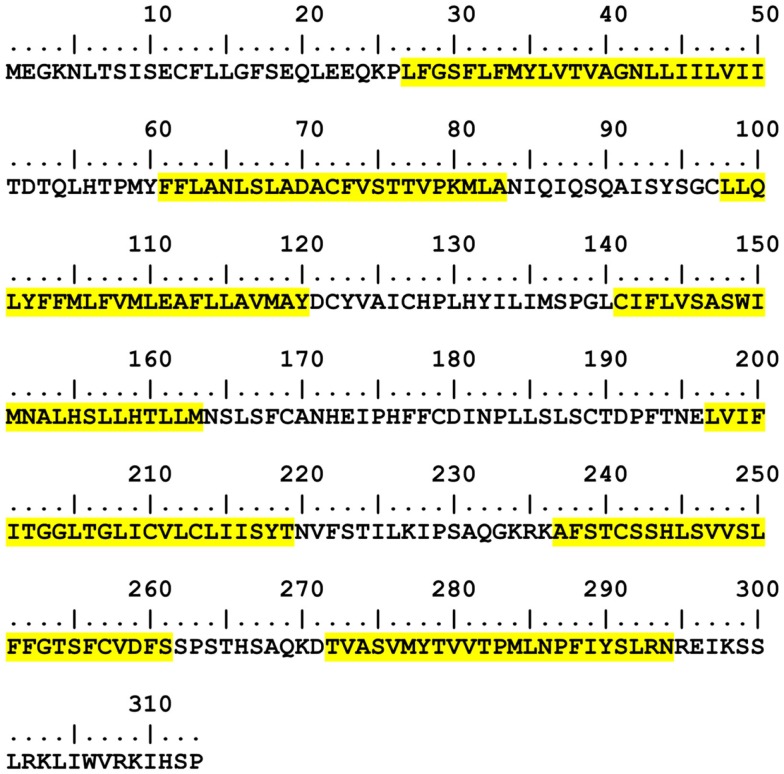
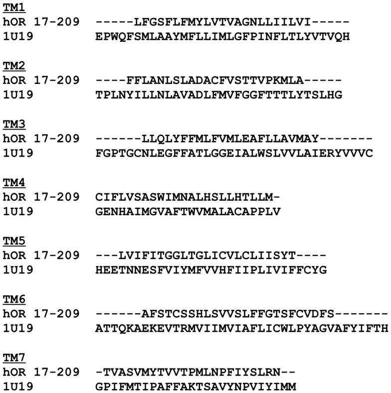
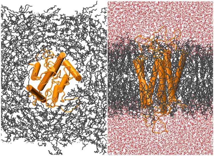
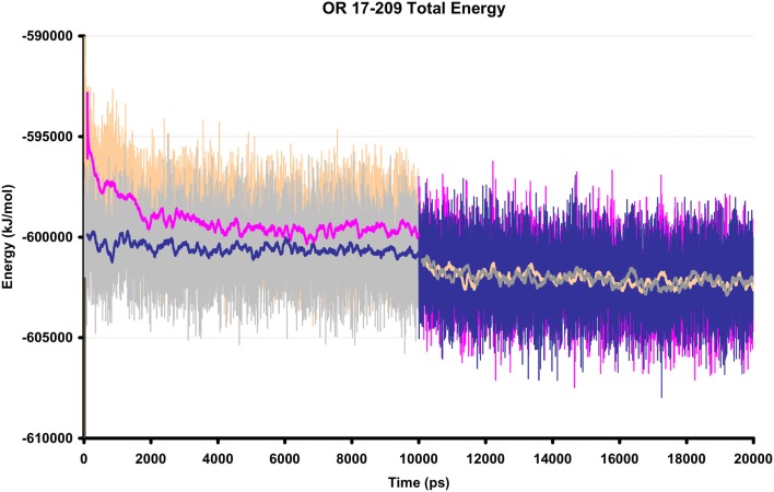
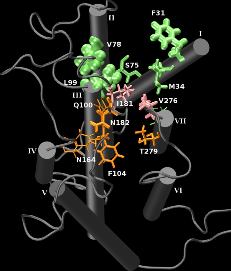
Olfactory receptors (ORs) are a type of GTP-binding protein-coupled receptor (GPCR). These receptors are responsible for mediating the sense of smell through their interaction with odor ligands. OR-odorant interactions marks the first step in the process that leads to olfaction. Computational studies on model OR structures can generate focused and novel hypotheses for further bench investigation by providing a view of these interactions at the molecular level beyond inferences that are drawn merely from static docking. Here we have shown the specific advantages of simulating the dynamic environment associated with OR-odorant interactions. We present a rigorous protocol which ranges from the creation of a computationally derived model of an olfactory receptor to simulating the interactions between an OR and an odorant molecule. Given the ubiquitous occurrence of GPCRs in the membranes of cells, we anticipate that our OR-developed methodology will serve as a model for the computational structural biology of all GPCRs.

Keywords: GPCR; ligand binding; lipid bilayer; molecular dynamics; olfactory receptor; protein modeling.


Figures

Figure 1
Figure 1
OR and GPCR end-to-end modeling and simulation flow diagram.
Figure 2
Figure 2
hOR17-209 TM prediction results from TMHMM. Predicted locations and lengths of transmembrane helices are highlighted in yellow. The non-highlighted regions are modeled as free-standing loops.
Figure 3
Figure 3
hOR17-209 TM prediction results and structural alignments against the TMs in Bovine Rhodopsin (PDB: 1U19).
Figure 4
Figure 4
hOR17-209 docked with its activating ligand, isopentyl acetate using GRAMM. The loops from the receptor model were removed prior to docking, and GRAMM was configured to provide 100 docked conformations. The physiologically relevant (extracellular and within the hydrophilic core) clusters are shown in blue and the discarded conformations are shown in red. One conformation from the blue cluster is selected for molecular dynamics simulation. (A) Longitudinal view. (B) Extracellular cross-section view.
Figure 5
Figure 5
hOR17-209 is embedded into the lipid bilayer-solvent complex with g_membed. The receptor, lipids, and waters are colored orange, gray, and red, respectively.
Figure 6
Figure 6
The total energy of two hOR 17-209 simulation systems, with and without docked ligand. The orange/pink regions show the total energy of the ligand-bound system and the gray/blue regions reflect the total energy of the unbound system during equilibration (first 10 ns) and production MD (final 10 ns). The solid lines are 200 ps moving averages. During production MD, no significant fluctuations are observed in the energy profiles.
Figure 7
Figure 7
A graphical representation of the interaction frequency between the carbonyl oxygen of isoamyl acetate and hOR 17-209 through 10 ns of MD. Green represents the primary binding pocket and orange represents a second binding pocket which is sampled by the ligand for 2 ns. Pink residues line the saddle point between the two regions. The larger the residue’s appearance, the higher the interaction frequency. The N- and C-termini were removed for clarity.

Similar articles

Cited by

References

    1. Allen W. J., Lemkul J. A., Bevan D. R. (2009). Gridmat-md: a grid-based membrane analysis tool for use with molecular dynamics. J. Comput. Chem. 30, 1952–195810.1002/jcc.21172 - DOI - PubMed
    1. Arora N., Jayaram B. (1996). Strength of hydrogen bonds in α helices. J. Comput. Chem. 18, 1245–125210.1002/(SICI)1096-987X(19970715)18:9<1245::AID-JCC12>3.0.CO;2-4 - DOI
    1. Buck L., Axel R. (1991). A novel multigene family may encode odorant receptors: a molecular basis for odor recognition. Cell 65, 175–18710.1016/0092-8674(91)90418-X - DOI - PubMed
    1. Crasto C. (2010). Hydrophobicity profiles in G protein-coupled receptor transmembrane helical domains. J. Receptor Ligand Channel Res. 3, 123–133 - PMC - PubMed
    1. Dong X., Han S. K., Zylka M. J., Simon M. I., Anderson D. J. (2001). A diverse family of GPCRs expressed in specific subsets of nociceptive sensory neurons. Cell 106, 619–63210.1016/S0092-8674(01)00483-4 - DOI - PubMed

LinkOut - more resources