

doi: 10.4070/kcj.2012.42.4.281.
Epub 2012 Apr 26.
A rare case of aortic valve myxoma: easy to confuse with papillary fibroelastoma
Affiliations
- PMID: 22563343
- PMCID: PMC3341427
- DOI: 10.4070/kcj.2012.42.4.281
Item in Clipboard
A rare case of aortic valve myxoma: easy to confuse with papillary fibroelastoma
Korean Circ J.
2012 Apr.
Abstract
Myxoma of the aortic valve is an exceedingly uncommon condition. In this article, we report the case of a 72-year-old man with myxoma arising from the aortic valve. We extirpated the mass and repaired the aortic valve with the patient under cardiopulmonary bypass. The postoperative course was uneventful. Histological examination confirmed that the mass was a myxoma.
Keywords: Aorta valve; Myxoma.
Conflict of interest statement
The authors have no financial conflicts of interest.
Figures
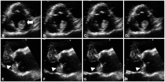
Echogenic mass in two-dimensional transthoracic echocardiography. This is serial images of cardiac tumor during the cardiac cycle. A-D were systole phase and E-H were diastole phase. A myxoma is seen on the aortic valve. A small spherical mass which shows relatively round surface with central necrosis was attached to the cusp of non coronary (A, arrow) and was highly mobile without pedicle. This tumor originated from the ventricular surface of the semi-lunar valve. It is attached to the leaflet of non coronary cusp (E-H, arrow head).
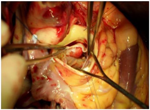
Intra-operation view. After aortic valve opening, photograph shows an ovoid mass attached below non coronary cusp.
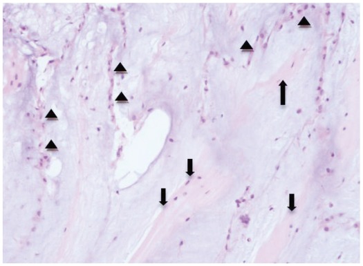
Histologic image, H-E stain (×200). Tumor consisted of gray purplish myxoid stroma with slender long spindle cells (arrow heads). Fibrocollagenous band portion of aortic valve is noted in the myxoma stroma (arrow).
References
LinkOut - more resources
Full Text Sources
