. 2012;7(4):e33905.
doi: 10.1371/journal.pone.0033905. Epub 2012 Apr 26.

Bloom's syndrome and PICH helicases cooperate with topoisomerase IIα in centromere disjunction before anaphase

Affiliations

Bloom's syndrome and PICH helicases cooperate with topoisomerase IIα in centromere disjunction before anaphase

Sébastien Rouzeau et al. PLoS One. 2012.

Abstract

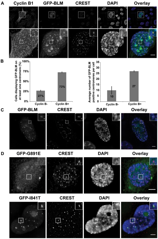
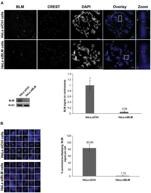
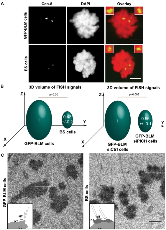
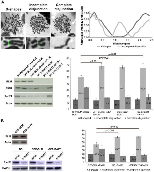
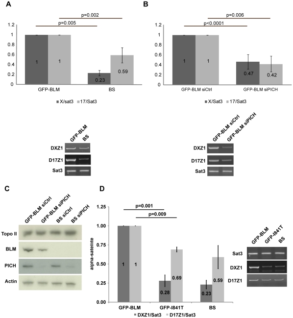
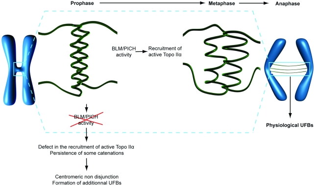
Centromeres are specialized chromosome domains that control chromosome segregation during mitosis, but little is known about the mechanisms underlying the maintenance of their integrity. Centromeric ultrafine anaphase bridges are physiological DNA structures thought to contain unresolved DNA catenations between the centromeres separating during anaphase. BLM and PICH helicases colocalize at these ultrafine anaphase bridges and promote their resolution. As PICH is detectable at centromeres from prometaphase onwards, we hypothesized that BLM might also be located at centromeres and that the two proteins might cooperate to resolve DNA catenations before the onset of anaphase. Using immunofluorescence analyses, we demonstrated the recruitment of BLM to centromeres from G2 phase to mitosis. With a combination of fluorescence in situ hybridization, electron microscopy, RNA interference, chromosome spreads and chromatin immunoprecipitation, we showed that both BLM-deficient and PICH-deficient prometaphase cells displayed changes in centromere structure. These cells also had a higher frequency of centromeric non disjunction in the absence of cohesin, suggesting the persistence of catenations. Both proteins were required for the correct recruitment to the centromere of active topoisomerase IIα, an enzyme specialized in the catenation/decatenation process. These observations reveal the existence of a functional relationship between BLM, PICH and topoisomerase IIα in the centromere decatenation process. They indicate that the higher frequency of centromeric ultrafine anaphase bridges in BLM-deficient cells and in cells treated with topoisomerase IIα inhibitors is probably due not only to unresolved physiological ultrafine anaphase bridges, but also to newly formed ultrafine anaphase bridges. We suggest that BLM and PICH cooperate in rendering centromeric catenates accessible to topoisomerase IIα, thereby facilitating correct centromere disjunction and preventing the formation of supernumerary centromeric ultrafine anaphase bridges.


Conflict of interest statement

Competing Interests: The authors have declared that no competing interests exist.

Figures

Figure 1
Figure 1. GFP-BLM localizes to centromeres in G2/prophase cells.
(A) Localization of GFP-BLM (green) to centromeres in late G2 cells. Nuclei were visualized by DAPI staining (blue). G2 cells were stained with antibodies against cyclin B1. Cells were stained with CREST serum to visualize centromeres (red). Lower panels are the magnification of the corresponding upper panels. Scale bar = 5 µm. (B) Two hundred cells with GFP-BLM-positive centromeres were analyzed by staining for cyclin B1 (cyclin B- or cyclin B+). The percentage of cells in each category is indicated (left panel), together with the mean number of GFP-BLM-positive centromeres per cell (right panel). Bars indicate the standard deviation (SD). (C) Localization of GFP-BLM (green) to centromeres (red) in prophase cells. The nucleus and centromeres were visualized as in A. Scale bar = 5 µm. (D) Localization of GFP-BLM (green) with an inactive helicase domain (GFP-I841T) or with both the helicase and DNA-binding domains inactivated (GFP-G891E). Nuclei and centromeres were visualized as in A. Scale bar = 5 µm.
Figure 2
Figure 2. BLM localizes to centromeres in mitotic cells.
(A) Endogenous BLM localizes to centromeres in HeLa cells. Wide-field microscopy after immunofluorescence staining on chromosome spreads obtained by cytocentrifugation of BLM siRNA-transfected or control siRNA-transfected HeLa S3 cells. Staining is shown for BLM (green), CREST (red) and chromosomes (blue). Single chromosome magnifications are shown (upper panels). Proteins levels were assessed by immunoblotting, probing the same membrane with anti-BLM (ab-476) antibody and then with anti-β actin antibody, as a loading control (lower left panel). Quantification of centromeric fluorescence signals for BLM (normalized according to the CREST signal) for a total of 20 centromeres in control cells (siCtrl) and 24 centromeres in BLM-depleted cells (siBLM) from two independent experiments, demonstrate the specificity of ab476 BLM antibodies (lower right panel). Data are means and SD normalized with respect to controls. Scale bar = 5 µm. (B) Centromeres detected with CREST serum (red) from all the chromosomes of HeLa siCtrl cells and HeLa siBLM cells from Figure 2A were analyzed for BLM signals (green). Chromosomes were visualized by DAPI staining (blue) (left panels). The same analysis was performed on a total of 9 HeLa siCtrl cells and 9 HeLa siBLM cells from two independent experiments: the percentage of centromeres giving a BLM signal is shown (right panel).
Figure 3
Figure 3. Structural defects at the centromeres in BLM-and PICH-deficient cells.
(A) FISH with the CEN-8 probe (green) on metaphase BS and GFP-BLM cells. Chromosomes are visualized by DAPI staining (red). Bar = 5 µm. (B) Comparison of the volume of the centromeric FISH signal detected on chromosomes 8 from GFP-BLM (defined as 1) and BS cells (left panel) and from GFP-BLM cells with (siPICH) and without (siCtrl) PICH downregulation (defined as 1) (right panel). We analyzed 45 metaphase cells from three independent experiments for each cell line. (C) GFP-BLM cells (left panel) and BS cells (right panel) were processed for electron microscopy. Scale bar = 1 µm. Inset: schematic diagram of the regions of interest (white squares) (MT: microtubules, KT: kinetochore outer plate and CC: centromeric chromatin). Eight centromeres from two independent experiments were analyzed.
Figure 4
Figure 4. BLM-deficient cells and PICH-downregulated display non disjunction of centromeres and impaired recruitment of active Topo IIα to centromeres.
(A) GFP-BLM and BS cells were transfected for 72 hours with Rad21 siRNAs and transfected either with control siRNAs or with PICH siRNAs. BLM, PICH and Rad21 proteins levels were assessed by immunoblotting, probing the same membrane with anti-BLM (ab-476), anti-PICH and anti-Rad21 antibodies and with anti-β actin antibody, as a loading control (lower left panel). Chromosome spreads were performed and sorted on the basis of their phenotype: X-shapes, incomplete disjunction or complete disjunction. The scale bars indicate 5 µm (upper left panel). This classification is based on the intensity profiles of centromeres (upper right panel). We analyzed 500 spreads from three independent experiments for each cell line. The frequency of each phenotype, in each of the three cell lines, is shown in the histogram (lower right panel). Bars represent SD. (B) GFP-BLM, BS and GFP-I841T cells were transfected for 72 hours with Rad21 siRNAs and the same experiments as in (A) (right panels) were carried out. We checked the levels of BLM and Rad21 proteins by western blotting (left panels).
Figure 5
Figure 5. PICH and BLM are required for the recruitment of Topo IIα to centromeres (A) ChIP-PCR analysis of Topo IIα recruitment to pericentromeric region satellite 3 (Sat3) or to the centromeric DNA of chromosomes X (DXZ1) or 17 (D17Z1) was performed in GFP-BLM and BS cells (lower panels).
The histogram shows relative enrichment in DXZ1 (DXZ1/Sat3) or D17Z1 (D17Z1/Sat3) sequences for active Topo IIα in GFP-BLM (defined as 1) and in BS cells (upper panels). The results presented are from three independent experiments. Error bars indicate the SD. (B) We performed the same experiments as in (A), with GFP-BLM cells transfected for 72 hours with control siRNAs or PICH siRNAs. (C) The amount of Topo II was evaluated in each cell line by western blotting. (D) We performed the same experiments as in (A), with GFP-BLM, GFP-I841T and BS cells.
Figure 6
Figure 6. Model of PICH/BLM-dependent decatenation of centromeric DNA.
We propose that the combined action of BLM and PICH promotes the organization of centromeric chromatin, thereby rendering some centromeric catenates accessible to Topo IIα. In the absence of BLM or PICH, a defect in the recruitment of active Topo IIα results in the persistence of some catenations leading to centromeric non disjunction and the formation of additional UFBs.

Similar articles

Cited by

References

    1. Carroll CW, Straight AF. Centromere formation: from epigenetics to self-assembly. Trends Cell Biol. 2006;16:70–78. - PubMed
    1. Cleveland DW, Mao Y, Sullivan KF. Centromeres and kinetochores: from epigenetics to mitotic checkpoint signaling. Cell. 2003;112:407–421. - PubMed
    1. Yuen KW, Montpetit B, Hieter P. The kinetochore and cancer: what’s the connection? Curr Opin Cell Biol. 2005;17:576–582. - PubMed
    1. Baumann C, Korner R, Hofmann K, Nigg EA. PICH, a centromere-associated SNF2 family ATPase, is regulated by Plk1 and required for the spindle checkpoint. Cell. 2007;128:101–114. - PubMed
    1. Chan KL, North PS, Hickson ID. BLM is required for faithful chromosome segregation and its localization defines a class of ultrafine anaphase bridges. EMBO J. 2007;26:3397–3409. - PMC - PubMed

Publication types

MeSH terms