. 2012;7(4):e35090.
doi: 10.1371/journal.pone.0035090. Epub 2012 Apr 26.

Effect of synthetic aβ peptide oligomers and fluorinated solvents on Kv1.3 channel properties and membrane conductance

Affiliations

Effect of synthetic aβ peptide oligomers and fluorinated solvents on Kv1.3 channel properties and membrane conductance

Maria I Lioudyno et al. PLoS One. 2012.

Abstract

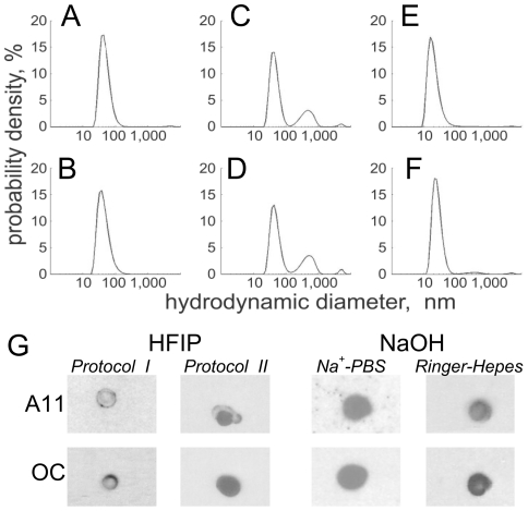
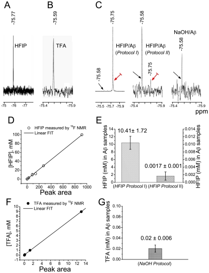
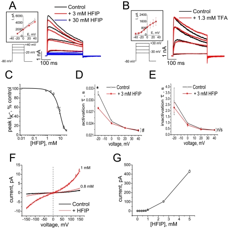
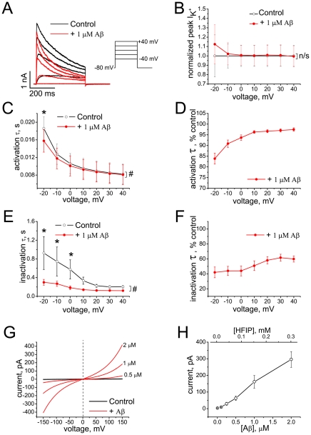
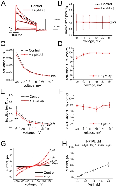
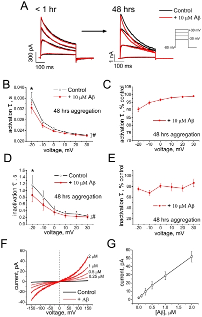
The impact of synthetic amyloid β (1-42) (Aβ(1-42)) oligomers on biophysical properties of voltage-gated potassium channels Kv 1.3 and lipid bilayer membranes (BLMs) was quantified for protocols using hexafluoroisopropanol (HFIP) or sodium hydroxide (NaOH) as solvents prior to initiating the oligomer formation. Regardless of the solvent used Aβ(1-42) samples contained oligomers that reacted with the conformation-specific antibodies A11 and OC and had similar size distributions as determined by dynamic light scattering. Patch-clamp recordings of the potassium currents showed that synthetic Aβ(1-42) oligomers accelerate the activation and inactivation kinetics of Kv 1.3 current with no significant effect on current amplitude. In contrast to oligomeric samples, freshly prepared, presumably monomeric, Aβ(1-42) solutions had no effect on Kv 1.3 channel properties. Aβ(1-42) oligomers had no effect on the steady-state current (at -80 mV) recorded from Kv 1.3-expressing cells but increased the conductance of artificial BLMs in a dose-dependent fashion. Formation of amyloid channels, however, was not observed due to conditions of the experiments. To exclude the effects of HFIP (used to dissolve lyophilized Aβ(1-42) peptide), and trifluoroacetic acid (TFA) (used during Aβ(1-42) synthesis), we determined concentrations of these fluorinated compounds in the stock Aβ(1-42) solutions by (19)F NMR. After extensive evaporation, the concentration of HFIP in the 100× stock Aβ(1-42) solutions was ∼1.7 μM. The concentration of residual TFA in the 70× stock Aβ(1-42) solutions was ∼20 μM. Even at the stock concentrations neither HFIP nor TFA alone had any effect on potassium currents or BLMs. The Aβ(1-42) oligomers prepared with HFIP as solvent, however, were more potent in the electrophysiological tests, suggesting that fluorinated compounds, such as HFIP or structurally-related inhalational anesthetics, may affect Aβ(1-42) aggregation and potentially enhance ability of oligomers to modulate voltage-gated ion channels and biological membrane properties.


Conflict of interest statement

Competing Interests: The authors have declared that no competing interests exist.

Figures

Figure 1
Figure 1. Size and conformation of Aβ1–42 aggregates.
(A–F) Size distributions of Aβ1–42 aggregates at different time points, as determined with DLS. (A–D) Aβ1–42 samples prepared with HFIP: (A, B: protocol I; C, D: protocol II) (E, F) Aβ1–42 samples prepared with NaOH and incubated at pH = 7. A, C, F show samples incubated for 48 h and B, D, F show samples incubated for 4 d at room temperature. Data taken at T = 25±0.1°C. (G) Dot blots probed with A11 and OC antibodies show that both A11-positive and OC-positive material is contained in the Aβ1–42 samples after 4 days of incubation. Representative blots are shown (n≥2 for each condition).
Figure 2
Figure 2. Quantification of HFIP and TFA in aqueous solutions by 19F NMR.
(A) Aβ-free aqueous solution spiked with 0.1 mM HFIP. (B) Aβ-free aqueous solution spiked with 0.13 mM TFA. (C) 19F NMR spectra of Aβ1–42 oligomer samples prepared using HFIP protocol I, HFIP protocol II, and the NaOH protocol. The signal amplification differs greatly between spectra as indicated by the different noise levels. The concentrations of Aβ1–42 and HFIP prior to evaporation were 70 μM and 1.2 M. Black and red arrows indicate peaks originating from residual TFA and HFIP, respectively. (D, F) Calibration standards generated by integrating the area under the 19F peaks obtained from samples with known HFIP (D) or TFA (F) concentrations. The lines correspond to the best fits through the origin (R 2 = 0.999 for both fits). (E) HFIP concentrations ± S.E.M. in stock Aβ1–42 samples prepared according to HFIP protocols I (n = 6) and II (n = 7). (G) TFA concentration ± S.E.M. in stock Aβ1–42 samples prepared according to NaOH protocol (n = 2).
Figure 3
Figure 3. HFIP and TFA effects on Kv1.3 channel currents and BLM conductance.
(A) Representative current traces before (black) and after (red and blue) application of HFIP (n = 5 cells). (B) Representative current traces before (black) and after (red) application of TFA (n = 3 cells). The current-voltage relations before and after application of HFIP (A) or TFA (B) are shown in the inserts. (C) Dose-response curve for the HFIP effect on peak K+ currents, evoked by depolarizing steps to +40 mV. IC50 = 5.2±3.4 mM from fitting the data (mean ± S.E.M., n = 5 cells for each concentration) to a Boltzmann function. (D) Activation of Kv 1.3 current is accelerated by HFIP (Data shown as mean ± S.E.M., n = 3 cells). The effect of HFIP on the activation time constant was significant (F = 50.6; #P = 0.01, Two-way RM-ANOVA) with significant interaction between FactorA (treatment) and FactorB (voltage) (F = 155), and by Pairwise Comparisons at −20 mV (*P = 7.9969×10⁻⁶, Tukey test). (E) The inactivation kinetics of Kv 1.3 current are not significantly affected by HFIP (mean ± S.E.M., n = 3 cells, #P = 0.36369, Two-way RM-ANOVA). (F) Representative I/V curves recorded on DOPC/DOPE BLMs in the presence of HFIP. (G) Dose-dependence of HFIP-induced currents through BLMs at +150 mV (mean ± S.E.M. for n = 7).
Figure 4
Figure 4. Effect of Aβ1–42 oligomers (HFIP protocol I) on Kv 1.3 currents and on BLM conductance.
(A) Representative K+ currents evoked by depolarizing voltage steps from holding potential of −80 mV before (black) and after (red) application of Aβ1–42 oligomers. (B) Peak K+ currents normalized to mean control values at different voltages before (black) and after application of Aβ1–42 oligomers (red). Data are shown as mean ± S.E.M. (n = 6 cells). HFIP had no significant (n/s) effect on the peak current (F = 0.17; P = 0.69, Two-way RM-ANOVA). (C–F) Activation and inactivation kinetics of K+ currents before (black) and after application of Aβ1–42 oligomers (red) shown in absolute (C, E) and normalized (D, F) values of time constants at different voltages (mean ± S.E.M., n = 6 cells). The effect of Aβ on the activation time constant was significant in Tests of Within-Subjects Effects (F = 46.8; #P = 4.7×10⁻⁴, Two-way RM-ANOVA), with significant interaction between FactorA (treatment) and FactorB (voltage) (F = 25.9), and by Pairwise Comparisons at −20 mV (*P = 2.08×10⁻⁴, Tukey test). The effect of Aβ on the inactivation time constant was also significant in Tests of Within-Subjects Effects (F = 8.1; #P = 0.04, Two-way RM-ANOVA), and by Pairwise Comparisons at −20 mV, −10 and 0 mV (*P<0.05; Tukey test). (G) Representative I/V curves recorded on DOPC/DOPE BLMs before (black) and after (red) application of Aβ1–42 oligomers. (H) Dose-dependence of Aβ1–42-induced currents at +150 mV across BLMs (mean ± S.E.M., n = 11 experiments, out of a total of 16, in which the effect was observed). HFIP concentrations estimated from 19F NMR spectra of the Aβ1–42 stock solutions are shown on the top axis.
Figure 5
Figure 5. Effects of Aβ1–42 oligomers (HFIP protocol II) on Kv 1.3 currents and on BLM conductance.
(A) Representative K+ currents evoked by depolarizing voltage steps from the holding potential of −80 mV before (black) and after (red) application of Aβ1–42 oligomers. (B) Peak K+ currents normalized to mean control values at different voltages after application of Aβ1–42 oligomers. The differences in peak current amplitude before and after application of Aβ are not significant (F = 3.9; P = 0.08 two-way RM-ANOVA). (C–F) Activation and inactivation kinetics of K+ currents before (black) and after application of Aβ1–42 oligomers (red) shown in absolute (C, E) and normalized (D, F) values of time constants at different voltages (mean ± S.E.M., n = 4 cells). The effect of Aβ on the activation time constant was not significant in Tests of Within-Subjects Effects (F = 4.4; #P = 0.07, Two-way RM-ANOVA). ANOVA analysis also revealed no significant effect of Aβ on the inactivation time constant (F = 8.7; #P = 0.05, Two-way RM-ANOVA), however two-way paired t-Test, (++P<0.05) showed significant differences between mean time constant measured before and after treatment with Aβ, revealing the trend. (G) Representative I/V curves recorded on DOPC/DOPE BLMs before (black) and after (red) application of Aβ1–42 oligomers. (H) Dose-dependence of Aβ-induced currents at +150 mV across BLMs (mean ± S.E.M., n = 7 experiments, out of a total of 9, in which the effect was observed).
Figure 6
Figure 6. Effect of HFIP-free Aβ1–42 oligomers (NaOH protocol) on Kv 1.3 current and on BLM conductance.
(A) Representative K+ currents evoked by depolarizing voltage steps from the holding potential of −80 mV before (black) and after (red) Aβ1–42 application. Note, that Aβ1–42 samples aggregated for less than 1 hr and presumably contained monomeric peptide, had no effect on K+ current (A, left), whereas samples aggregated for 48 hrs produced characteristic effect on K+ current kinetics (A, right). (B–E) Activation and inactivation kinetics of K+ currents before (black) and after application of Aβ1–42 oligomers (red) shown in absolute (B, D) and normalized (C, E) values of time constants at different voltages (mean ± S.E.M., n = 4 cells). The effect of Aβ on the activation time constant was significant in Tests of Within-Subjects Effects (F = 34.5; #P = 0.009, Two-way RM-ANOVA) with significant interaction between FactorA (treatment) and FactorB (voltage) (F = 38.03), and by Pairwise Comparisons at −20 mV (*P = 0.006, Tukey test). The effect of Aβ on the inactivation time constant was also significant in Tests of Within-Subjects Effects (F = 19.1; #P = 0.022, Two-way RM-ANOVA) with significant interaction between FactorA (treatment) and FactorB (voltage) (F = 7.1), and by Pairwise Comparisons at −20 mV (*P = 0.016, Tukey test). (F) Representative I/V curves recorded on DOPC/DOPE BLMs before (black) and after (red) application of Aβ1–42 oligomers. (G) Dose-dependence of Aβ-induced currents at +150 mV across BLMs (mean ± S.E.M., n = 5 experiments, out of a total of 12, in which the effect was observed).

Similar articles

Cited by

References

    1. Bertram L, Lill CM, Tanzi RE. The Genetics of Alzheimer Disease: Back to the Future. Neuron. 2010;68:270–281. - PubMed
    1. Chouliaras L, Rutten BPF, Kenis G, Peerbooms O, Visser PJ, et al. Epigenetic regulation in the pathophysiology of Alzheimer's disease. Prog Neurobiol. 2010;90:498–510. - PubMed
    1. Esteban JA. Living with the enemy: a physiological role for the [beta]-amyloid peptide. Trends Neurosci. 2004;27:1–3. - PubMed
    1. Parihar MS, Brewer GJ. Amyloid-ß as a modulator of synaptic plasticity. J Alzheimers Dis. 2010;22:741–763. - PMC - PubMed
    1. Ramsden M, Henderson Z, Pearson HA. Modulation of Ca2+ channel currents in primary cultures of rat cortical neurones by amyloid [beta] protein (1–40) is dependent on solubility status. Brain Res. 2002;956:254–261. - PubMed

Publication types