. 2012;7(4):e35642.
doi: 10.1371/journal.pone.0035642. Epub 2012 Apr 26.

The dopamine D2 receptor gene in lamprey, its expression in the striatum and cellular effects of D2 receptor activation

Affiliations

The dopamine D2 receptor gene in lamprey, its expression in the striatum and cellular effects of D2 receptor activation

Brita Robertson et al. PLoS One. 2012.

Abstract

All basal ganglia subnuclei have recently been identified in lampreys, the phylogenetically oldest group of vertebrates. Furthermore, the interconnectivity of these nuclei is similar to mammals and tyrosine hydroxylase-positive (dopaminergic) fibers have been detected within the input layer, the striatum. Striatal processing is critically dependent on the interplay with the dopamine system, and we explore here whether D2 receptors are expressed in the lamprey striatum and their potential role. We have identified a cDNA encoding the dopamine D2 receptor from the lamprey brain and the deduced protein sequence showed close phylogenetic relationship with other vertebrate D2 receptors, and an almost 100% identity within the transmembrane domains containing the amino acids essential for dopamine binding. There was a strong and distinct expression of D2 receptor mRNA in a subpopulation of striatal neurons, and in the same region tyrosine hydroxylase-immunoreactive synaptic terminals were identified at the ultrastructural level. The synaptic incidence of tyrosine hydroxylase-immunoreactive boutons was highest in a region ventrolateral to the compact layer of striatal neurons, a region where most striatal dendrites arborise. Application of a D2 receptor agonist modulates striatal neurons by causing a reduced spike discharge and a diminished post-inhibitory rebound. We conclude that the D2 receptor gene had already evolved in the earliest group of vertebrates, cyclostomes, when they diverged from the main vertebrate line of evolution (560 mya), and that it is expressed in striatum where it exerts similar cellular effects to that in other vertebrates. These results together with our previous published data (Stephenson-Jones et al. 2011, 2012) further emphasize the high degree of conservation of the basal ganglia, also with regard to the indirect loop, and its role as a basic mechanism for action selection in all vertebrates.


Conflict of interest statement

Competing Interests: The authors have declared that no competing interests exist.

Figures

Figure 1
Figure 1. The lamprey dopamine D2 receptor.
A. A schematic drawing of the lamprey dopamine D2 receptor nucleotide sequence showing the conserved regions (black) at the 5′ and 3′ ends, respectively, and the site from which the riboprobe (660 bp, red) was made. B. The seven transmembrane domains (TM-helix) are all located at the conserved parts of the dopamine D2 receptor. The deduced amino acid sequence of the receptor shows that it belongs to the GPCR-superfamily.
Figure 2
Figure 2. Alignment of dopamine D2 receptor amino acid sequences.
Alignment of the deduced amino acid sequence from the lamprey dopamine D2 receptor with those from Homo sapiens, Mus musculus, Danio rerio and Anguilla anguilla. The seven transmembrane domains are indicated.
Figure 3
Figure 3. Phylogenetic analysis of vertebrate dopamine receptors of the D2-family.
The distant tree was built with the neighbor-joining algorithm from alignments of the D2, D3 and D4 receptor subtypes from several vertebrate species. Data were re-sampled by 1000 bootstrap replicates to determine confidence indices within the phylogenetic tree. The scale bar refers to a phylogenetic distance of 0.09 amino acid substitution per site. The different vertebrate classes are indicated by different colors (mammals, blue; birds, dark green; reptiles, light green; amphibians, brown; fish, red; cyclostomes, mauve). GenBank accession numbers of the sequences are: Homo sapiens D2, AAA52761; Macaca mulatta D2, XP_001085571; Bos taurus D2, DAA22356; Canis lupus familiaris D2, AAG34494; Mus musculus D2, NP_034207; Rattus norvegicus D2, NP_036679; Gallus gallus D2, NP_001106761; Meleagris gallopavo D2, AAD03818; Anoils carolinensis D2, XP_ 003217484; Xenopus tropicalis D2, XP_002937871; Xenopus laevis D2, CAA51412; Rana catesbeiana D2, BAI70438; Mugil cephalus D2, AAU87970; Oreochromis niloticus D2, AAU87971; Tetraodon nigroviridis D2, CAF97490 ; Oncorhynchus mykiss D2, CAC87873; Danio rerio D2a, AAN87174; Danio rerio D2b, AAP94011; Anguilla anguilla D2A, ABH06893; Anguilla anguilla D2B, ABH06894; Lampetra fluviatilis D2, ADO23655; Mus musculus D3, 2105315A; Rattus norvegicus D3, 1614344A; Canis lupus familiaris D3, XP_545106; Homo sapiens D3, 1705199A ; Bos taurus D3, NP_001179824; Carassius auratus D3, ABN70936; Danio rerio D3, AAN87173; Homo sapiens D4, 1709359A; Macaca mulatta D4, XP_001087197; Mus musculus D4, 2109259A; Rattus norvegicus D4, AAA18588; Danio rerio D4a, AAW80614; Danio rerio D4b, AAW80615; Danio rerio D4c, AAW80616.
Figure 4
Figure 4. Dopamine D2 receptor expression in the lamprey brain.
A. 35S-UTP-labeled D2 receptor riboprobe shows strong expression in the striatum. D2 receptor expression is also present in the dorsal and lateral pallium although less strong. Note the absence of receptor expression in the magnocellular preoptic nucleus (arrow). B. Nissl stained section from the striatum showing the site of receptor expression in A and C. C. DIG-labeled D2 receptor riboprobe expressed in a subpopulation of striatal neurons. D. DIG-labeled sense riboprobe showing lack of mRNA expression in the section adjacent to the one in Figure 5C. E. 35S-UTP-labeled D2 receptor riboprobe expression in the habenula, thalamus and hypothalamus. F. Schematic drawing of the lamprey brain indicating the habenula, thalamus and hypothalamus. G. 35S-UTP-labeled D2 receptor riboprobe shows strong expression in the mammillary area. H. Schematic drawing of the lamprey brain indicating the mammillary area. The pseudocoloring in A, E and G indicates signal intensity from low (black/blue) to high (pink/yellow). Abbreviation: DPal, dorsal pallium; Hb, habenula; Hyp, hypothalamus; LPal, lateral pallium; MAM, mammillary region; ot, optic tract; OT, optic tectum; PO, preoptic nucleus; Str, Striatum; Th, thalamus. Scale bars, A, B, E and G 500 µm; C and D 200 µm.
Figure 5
Figure 5. Striatal projection neurons targeting substantia nigra pars reticulata (SNr) do not express the D2 receptor.
A. Striatal neurons retrogradely labeled after injections of neurobiotin into the SNr (white arrows). B. DIG-labeled D2 receptor riboprobe expressed in a subpopulation of striatal cells (black arrow heads show examples of positive cells). C. Merged image showing the absence of overlap between retrogradely labeled cells and D2 receptor mRNA expression. Scale bars, 100 µm.
Figure 6
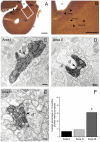
Figure 6. Tyrosine hydroxylase (TH) immunoreactivity in the lamprey striatum.
A and B. Light microscope photomicrographs showing the areas (I, II and III) of the striatum that were included in the electron microscope analysis. Note the slightly higher density of TH-immunoreactive fibers (some indicated by small arrows) in Areas I and III. C–E. Electron micrographs of TH-immunolabeled axonal profiles forming asymmetrical synapses (arrows) with dendritic shafts (d) in Area I (C), II (D) and III (E) of the lamprey striatum. Note the densely packed vesicles and the prominent postsynaptic densities. F. Quantitative analysis of synaptic incidence in the three areas of striatum. The histogram shows the average percentage of TH-immunolabeled profiles which form synaptic junctions (out of a total of 75 profiles per area, n = 3). Synaptic incidence in Area III was significantly higher when compared to Area I (x 2 test, p = 0.0003) and Area II (x 2 test, p = 0.0022). Abbreviations: vt, ventriculus medius telencephali; lpv, ventral part of the lateral pallium. Scale bars: A: 200 µm, B: 100 µm, C–E: 200 nm.
Figure 7
Figure 7. Modulation of striatal neurons by a dopamine D2 agonist.
A. Voltage responses of a striatal neuron to 1 s long hyperpolarizing and depolarizing current injections during control (A1), D2 agonist application (TNPA, A2) and washout (A3). B. The number of evoked action potentials is reduced upon application of TNPA, measured here at twice the threshold stimulation. C. The average number of PIR spikes is reduced by TNPA, measured after termination of five consecutive hyperpolarizations in between −100 and −80 mV. D. The total number of PIR spikes and the time until the spike are reduced by TNPA, measured as in C. E. Table of firing properties before and during TNPA. All values presented as mean ± SD and differences were significant for all measurements apart from the AP half-width (p<0.05, student's t-test). Abbreviations: AP, action potential; PIR, post-inhibitory rebound; TNPA, trihydroxy-N-propyl-noraporphine 123 hydrobromide hydrate.

Similar articles

Cited by

References

    1. Grillner S. Ion channels and locomotion. Science. 1997;278:1087–1088. - PubMed
    1. Graybiel AM. The basal ganglia and chunking of action repertoires. Neurobiol Learn Mem. 1998;70:119–136. - PubMed
    1. Redgrave P, Prescott TJ, Gurney K. The basal ganglia: a vertebrate solution to the selection problem? Neuroscience. 1999;89:1009–1023. - PubMed
    1. Hikosaka O, Takikawa Y, Kawagoe R. Role of the basal ganglia in the control of purposive saccadic eye movements. Physiol Rev. 2000;80:953–978. - PubMed
    1. Grillner S, Hellgren J, Menard A, Saitoh K, Wikstrom MA. Mechanisms for selection of basic motor programs–roles for the striatum and pallidum. Trends Neurosci. 2005;28:364–370. - PubMed

Publication types