. 2012;7(4):e35645.
doi: 10.1371/journal.pone.0035645. Epub 2012 Apr 26.

Zebrafish chemical screening reveals the impairment of dopaminergic neuronal survival by cardiac glycosides

Affiliations

Zebrafish chemical screening reveals the impairment of dopaminergic neuronal survival by cardiac glycosides

Yaping Sun et al. PLoS One. 2012.

Abstract

Parkinson's disease is a neurodegenerative disorder characterized by the prominent degeneration of dopaminergic (DA) neurons among other cell types. Here we report a first chemical screen of over 5,000 compounds in zebrafish, aimed at identifying small molecule modulators of DA neuron development or survival. We find that Neriifolin, a member of the cardiac glycoside family of compounds, impairs survival but not differentiation of both zebrafish and mammalian DA neurons. Cardiac glycosides are inhibitors of Na(+)/K(+) ATPase activity and widely used for treating heart disorders. Our data suggest that Neriifolin impairs DA neuronal survival by targeting the neuronal enriched Na(+)/K(+) ATPase α3 subunit (ATP1A3). Modulation of ionic homeostasis, knockdown of p53, or treatment with antioxidants protects DA neurons from Neriifolin-induced death. These results reveal a previously unknown effect of cardiac glycosides on DA neuronal survival and suggest that it is mediated through ATP1A3 inhibition, oxidative stress, and p53. They also elucidate potential approaches for counteracting the neurotoxicity of this valuable class of medications.


Conflict of interest statement

Competing Interests: The authors have declared that no competing interests exist.

Figures

Figure 1
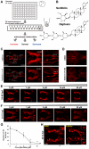
Figure 1. Zebrafish chemical screen identifies Neriifolin, a member of cardiac glycoside family, which disrupts the pattern of DA neurons in the ventral forebrain.
(A) Schematic diagram of the chemical screening platform, through which Neriifolin was identified as a hit that decreases ventral forebrain DA neurons. (B) Structure of two cardiac glycosides, Neriifolin and Digitoxin, both of which disrupt the pattern of VFB DA neurons. (C) Embryos treated with 10 µM Neriifolin showed a decrease of VFB DA neurons (middle panels), whereas the Sym NA neurons were normal (right panels). (D) Treatment with another cardiac glycoside, Digitoxin, similarly decreased VFB DA neurons but not Sym NA neurons. (E) Embryos treated with different concentrations of Neriifolin from 24 hpf to 48 hpf showed no obvious defect in the pattern of VFB DA neurons. (F) Embryos treated with different concentrations of Neriifolin from 24 hpf to 72 hpf displayed impaired DA neuron pattern in VFB. The dose response curve is shown in (G). (H) Embryos treated with Neriifolin from 48–72 hpf also showed deficit in VFB DA neurons: neuronal numbers in the control vs. treated embryo are 64 and 39 respectively. The insets show enlarged views of DA neurons, which reveal the presence of TH in the nucleus, indicating a loss of nuclear membrane integrity. OB, olfactory bulb; VFB, ventral forebrain; sym NA, sympathetic NA neurons; AAC NA, arch-associated NA; LC, locus coeruleus.
Figure 2
Figure 2. Human atp1a3 rescues DA neurons in Neriifolin-treated embryos.
(A) The expression pattern of atp1a3a in wild-type embryos at 48 hpf. (B) The schematic diagram of the plasmid constructs used for the rescue experiments in zebrafish embryos. (C) RT-PCR detection of the expression of human atp1a3 in zebrafish embryos after injection and heat shock. (D–E) Quantification (D) and representative images (E) of VFB DA neurons in 5 µM Neriifolin-treated embryos that express either GFP or human atp1a3. Data are the averages ± SEM from 9 embryos in a single experiment that was repeated twice with similar results.
Figure 3
Figure 3. Mechanisms and protective strategies for Neriifolin-induced DA neuronal death.
(A) Treatment with Neriifolin in the presence of high K+ in the medium significantly decreased DA neuronal loss compared to the treatment with Neriifolin alone. Data are the averages ± SEM from 6 embryos in a single experiment that was repeated twice with similar results. (B) Treatment with Neriifolin in the presence of low Na+ in the medium significantly decreased DA neuronal loss compared to the treatment with Neriifolin alone. Data are the averages ± SEM from 8 embryos in a single experiment that was repeated twice with similar results. (C–D) Treatment with either Quercetin (C) or Ascorbic Acid (D) significantly decreased DA neuronal death compared to treatment with Neriifolin alone. Data are the averages ± SEM from 8 embryos in a single experiment that was repeated twice with similar results.
Figure 4
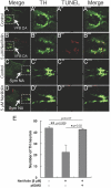
Figure 4. Neriifolin-induced DA neuronal death is apoptotic and requires p53.
(A–D′″) Low (A–D) and high (A′–D′″) magnification views of VFB DA neurons in control (A-A′″) vs Neriifolin-treated embryos (B-B′″), and sympathetic (Sym) NA neurons in control (C-C′″) vs Neriifolin-treated embryos (D-D′″). Ventral views of 60 hpf embryos were shown. Neriifolin treatment was carried out from 24 hpf to 60 hpf. (E) Injection of the p53-MO into embryos at one-cell stage protected DA neurons from cell death induced by Neriifolin. Data are the averages ± SEM from 6 embryos in a single experiment that was repeated twice with similar results.
Figure 5
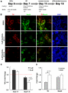
Figure 5. Neriifolin selectively impairs the survival of mammalian DA neurons derived from embryonic stem cells (ESCs).
(A) Schematic diagram of the protocol used to induce DA neuron differentiation from mouse ESCs. (B) Representative views from wells with different treatment, showing the percentage of TH+ neurons (TH, a marker of DA neurons, in red) or all neurons (NeuN, a neuronal marker, in green) among total cells (DAPI). Compared to DMSO control (upper panel), those cells treated with Neriifolin from Day 7 to 11 (middle panels) showed no decrease in the percentage of TH+ neurons and all neurons; but those cells treated with Neriifolin from day 11 to 14 (lower panels) showed significant less percentage of DA neurons. (C) Quantification of the percentage of TH+ neurons among total cells. Fold change of treatment vs DMSO control was shown. (D) Quantification of fold change of TH+ neurons and all neurons. In both (C) and (D), data are the averages ± SEM of triplicates from a single experiment that was repeated 3 times with similar results.
Figure 6
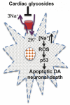
Figure 6. A schematic model showing the effect of cardiac glycosides on DA neuronal death.
We propose that inhibition of Na+/K+ ATPase activity leads to increased intracellular sodium, which triggers the production of reactive oxygen species (ROS) and activation of p53-mediated apoptotic cell death pathway. Future biochemical experiments are needed to further test this model.

Similar articles

Cited by

References

    1. Björklund A, Lindvall O. Dopamine-containing systems in the CNS. In: Björklund A, Hökfelt T, editors. Handbook of Chemical Neuroanatomy. Amsterdam: Elsevier; 1984. pp. 55–122.
    1. Goldstein DS, Eisenhofer G, McCarty R. Catecholamines: bridging basic science with clinical medicine; In: August JT, Anders MW, Murad F, Coyle JT, editors. San Diego, California: Academic Press; 1998.
    1. Chaudhuri KR, Schapira AH. Non-motor symptoms of Parkinson's disease: dopaminergic pathophysiology and treatment. Lancet Neurol. 2009;8:464–474. - PubMed
    1. Meissner WG, Frasier M, Gasser T, Goetz CG, Lozano A, et al. Priorities in Parkinson's disease research. Nat Rev Drug Discov. 2011;10:377–393. - PubMed
    1. Smeets WJAJ, Reiner A. Phylogeny and development of catecholamine systems in the CNS of vertebrates. Cambridge, England: Cambridge University Press; 1994.

Publication types

MeSH terms