. 2012;7(5):e35865.
doi: 10.1371/journal.pone.0035865. Epub 2012 May 1.

Rd9 is a naturally occurring mouse model of a common form of retinitis pigmentosa caused by mutations in RPGR-ORF15

Affiliations

Rd9 is a naturally occurring mouse model of a common form of retinitis pigmentosa caused by mutations in RPGR-ORF15

Debra A Thompson et al. PLoS One. 2012.

Abstract

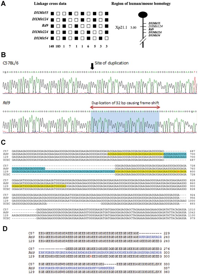
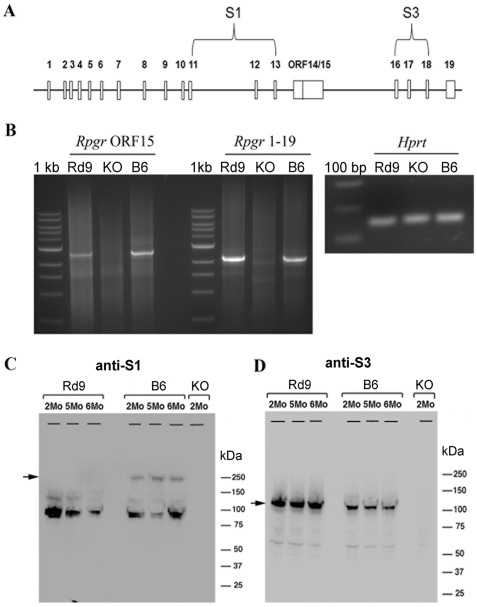
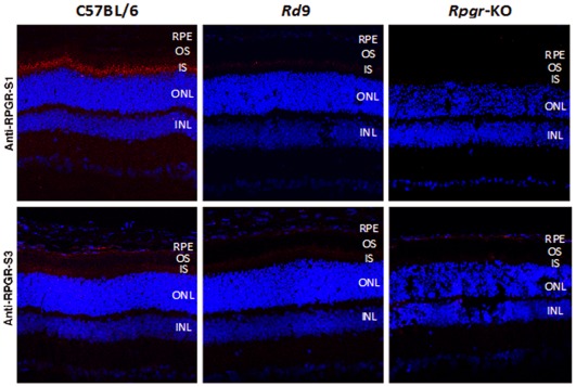
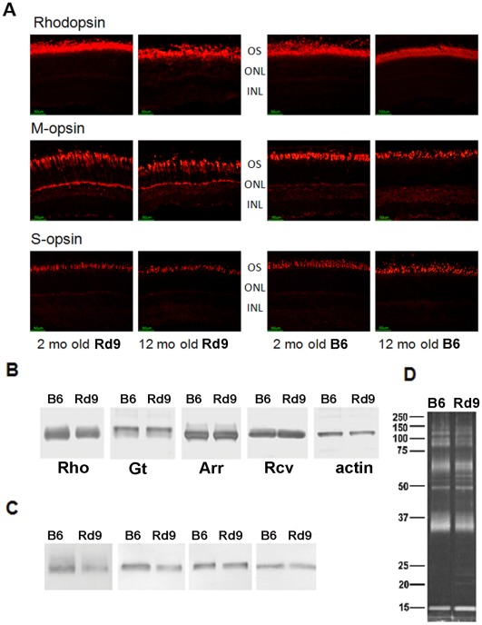
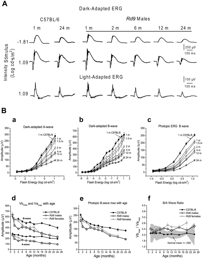
Animal models of human disease are an invaluable component of studies aimed at understanding disease pathogenesis and therapeutic possibilities. Mutations in the gene encoding retinitis pigmentosa GTPase regulator (RPGR) are the most common cause of X-linked retinitis pigmentosa (XLRP) and are estimated to cause 20% of all retinal dystrophy cases. A majority of RPGR mutations are present in ORF15, the purine-rich terminal exon of the predominant splice-variant expressed in retina. Here we describe the genetic and phenotypic characterization of the retinal degeneration 9 (Rd9) strain of mice, a naturally occurring animal model of XLRP. Rd9 mice were found to carry a 32-base-pair duplication within ORF15 that causes a shift in the reading frame that introduces a premature-stop codon. Rpgr ORF15 transcripts, but not protein, were detected in retinas from Rd9/Y male mice that exhibited retinal pathology, including pigment loss and slowly progressing decrease in outer nuclear layer thickness. The levels of rhodopsin and transducin in rod outer segments were also decreased, and M-cone opsin appeared mislocalized within cone photoreceptors. In addition, electroretinogram (ERG) a- and b-wave amplitudes of both Rd9/Y male and Rd9/Rd9 female mice showed moderate gradual reduction that continued to 24 months of age. The presence of multiple retinal features that correlate with findings in individuals with XLRP identifies Rd9 as a valuable model for use in gaining insight into ORF15-associated disease progression and pathogenesis, as well as accelerating the development and testing of therapeutic strategies for this common form of retinal dystrophy.


Conflict of interest statement

Competing Interests: The authors have declared that no competing interests exist.

Figures

Figure 1
Figure 1. Identification of the genetic defect on the X-chromosome in Rd9 mice.
A) Haplotype analysis showing the segregation patterns of Rd9 and flanking markers in (CAST/Rd9)F1 x B6 backcrosses generating 364 meioses. Each column represents the chromosome inherited by a group of backcross progeny, with black boxes representing BB homozygotes and white boxes denoting BC heterozygotes for a given locus. The number of offspring that inherited the haplotype of that region of the X Chromosome is listed at the bottom of each column. Also shown is the partial chromosome linkage map with the positions of Rd9 and its flanking markers. B) Chromatograms showing partial genomic sequence of Rpgr-ORF15 in B6 and Rd9 mice, showing the location of a 32 bp duplication in the Rd9 strain. The precise location is difficult to define because of the highly repetitive nature of the sequence. C) Comparison of Rpgr-ORF15 genomic sequences of B6, Rd9, and 129/SvJ mice determined by DNA sequence analysis and aligned using ClustalW2. The B6 genomic sequence of Rpgr-ORF15 from the UCSC genome browser is also included. Additional tandem duplications found in the Rd9 and 129/SvJ sequences are highlighted in yellow and blue. D) Alignment of Rpgr-ORF15 amino acid sequences derived by translation of the B6, Rd9, and 129/SvJ genomic sequences, and the sequence of B6 from the UCSC Genome Browser. The sequences were aligned using ClustalW2.
Figure 2
Figure 2. RT-PCR and immunoblot analysis of Rpgr expression in mutant and wild-type mice.
A) Schematic of the mouse Rpgr gene showing the locations of the ORF15 exon, the oligonucleotide primers for RT-PCR, and protein sequences used to generate the RPGR antibodies S1 and S3. B) RT-PCR was performed on total retinal RNA from two-month-old male mice using a common exon 1 primer and reverse primers for ORF15 or exon 19. The PCR products were resolved on a 1% agarose gel. RT-PCR of Hprt was used as a control for RNA recovery and loading. C) Western blots of retinal extracts from 2-6 month-old male B6 (wild type), Rd9, and Rpgr-KO mice probed with RPGR antibodies. The S1 antibody recognizes a sequence common to both Rpgr ORF15 and 1–19 variants. The S3 antibody recognizes only the protein corresponding to the 1–19 variant.
Figure 3
Figure 3. Immunohistochemical analysis of Rpgr in mutant and wild-type mice.
Cryosections of eyes from 2-month-old B6, Rd9, and Rpgr-KO male mice were probed with S1 antibody that recognizes a sequence common to both Rpgr ORF15 and 1–19 variants, and S3 antibody that recognizes a sequence unique to the 1–19 variant. Red shows Rpgr-specific staining; blue shows DAPI staining of nuclei.
Figure 4
Figure 4. Analysis of retinal histology in Rd9 and wild-type mice.
A) Photographs of plastic retina sections from Rd9/Y and B6 mice at the ages indicated. Morphometric analysis showing B) outer plus inner segment (OS+IS), and C) outer nuclear layer (ONL) thickness, in mice at 12 months-of-age. Thickness measurements were taken on sections parallel (superior/inferior) or orthogonal (nasal/temporal) to the vertical meridian of eyes from Rd9/Y (○) and B6 (▪) mice and plotted vs. distance from the optic nerve head, with standard deviations shown.
Figure 5
Figure 5. Rhodopsin and cone opsin expression in Rd9 and wild-type mice.
A) Immunohistochemical analysis of M-opsin, S-opsin, and rhodopsin in the superior retina in Rd9/Y and B6 mice at 2 and 12 months-of-age (red fluorescence labeling). M-cone opsin reactivity appears throughout both outer and inner segments in Rd9 mice at both ages. Rhodopsin reactivity is disturbed in Rd9 mice at 12 months-of-age. B) Western analysis of rhodopsin, transducin-alpha subunit, arrestin, and recoverin in blots of total eye retinal protein homogenates from Rd9 and B6 mice at 1 month-of-age. The band corresponding to monomeric rhodopsin is shown, and β-actin served as a loading control. C) Immunoblot analysis of rhodopsin, transducin, arrestin, and recoverin in ROS preparations from Rd9 and B6 mice. D) SyproRuby staining of ROS proteins separated by SDS-PAGE verified equivalent loading of Rd9 and wild-type protein (β-actin, Hprt, and Gapdh were not informative markers for ROS preparations). Representative results are shown (n = 3). Rhodopsin (Rho), transducin-alpha subunit (GT-α), arrestin (Arr), and recoverin (Rcv).
Figure 6
Figure 6. Electroretinogram (ERG) waveforms for Rd9 and wild-type mice.
A) Representative dark-adapted and light-adapted ERGs waveforms. For mice at 1 to 24 months-of-age, dark-adapted ERG recordings show the rod-mediated responses and maximal responses (rod plus cone), and light-adapted ERG recordings show the maximal cone-mediated responses. B) ERG intensity-response function V-Log I curves in Rd9 males (open symbols) at 1 (n =  10), 1.5 (n = 6), 2 (n = 13), 4 (n = 11), 12 (n = 11), and 24 months (n = 5) compared to 1-month-old B6 mice (filled squares). a) dark-adapted a-wave. b) dark-adapted b-wave. c) photopic b-wave. Natural history of maximum ERG amplitude (Mean ± SE) changes in Rd9/Y males, Rd9/Rd9 females, and B6 mice. d) Dark-adapted Vamax and Vbmax. e) Photopic Lmax. f) b/a wave ratio. Grey area represents the wild-type normal range.

Similar articles

Cited by

References

    1. Daiger SP, Bowne SJ, Sullivan LS. Perspective on genes and mutations causing retinitis pigmentosa. Arch Ophthalmol. 2007;125:151–158. - PMC - PubMed
    1. Fishman GA, Farber MD, Derlacki DJ. X-linked retinitis pigmentosa: Profile of clinical findings. Arch Ophthalmol. 1988;106:369–375. - PubMed
    1. Breuer DK, Yashar BM, Filippova E, Hiriyanna S, Lyons RH, et al. A comprehensive mutation analysis of RP2 and RPGR in a North American cohort of families with X-linked retinitis pigmentosa. Am J Hum Genet. 2002;70:1545–1554. - PMC - PubMed
    1. Shu X, Blac GC, Rice JM, Hart-Holden N, Jones A, et al. RPGR mutation analysis and disease: an update. Hum Mutat. 2007;28:322–328. - PubMed
    1. Vervoort R, Lennon A, Bird AC, Tulloch B, Axton R, et al. Mutational hot spot within a new RPGR exon in X-linked retinitis pigmentosa. Nat Genet. 2000;25:462–466. - PubMed

Publication types

MeSH terms