. 2012;7(5):e36337.
doi: 10.1371/journal.pone.0036337. Epub 2012 May 1.

Alternative splicing of Spg7, a gene involved in hereditary spastic paraplegia, encodes a variant of paraplegin targeted to the endoplasmic reticulum

Affiliations

Alternative splicing of Spg7, a gene involved in hereditary spastic paraplegia, encodes a variant of paraplegin targeted to the endoplasmic reticulum

Giuseppe Mancuso et al. PLoS One. 2012.

Abstract

Background: Hereditary spastic paraplegia defines a group of genetically heterogeneous diseases characterized by weakness and spasticity of the lower limbs owing to retrograde degeneration of corticospinal axons. One autosomal recessive form of the disease is caused by mutation in the SPG7 gene. Paraplegin, the product of SPG7, is a component of the m-AAA protease, a high molecular weight complex that resides in the mitochondrial inner membrane, and performs crucial quality control and biogenesis functions in mitochondria.

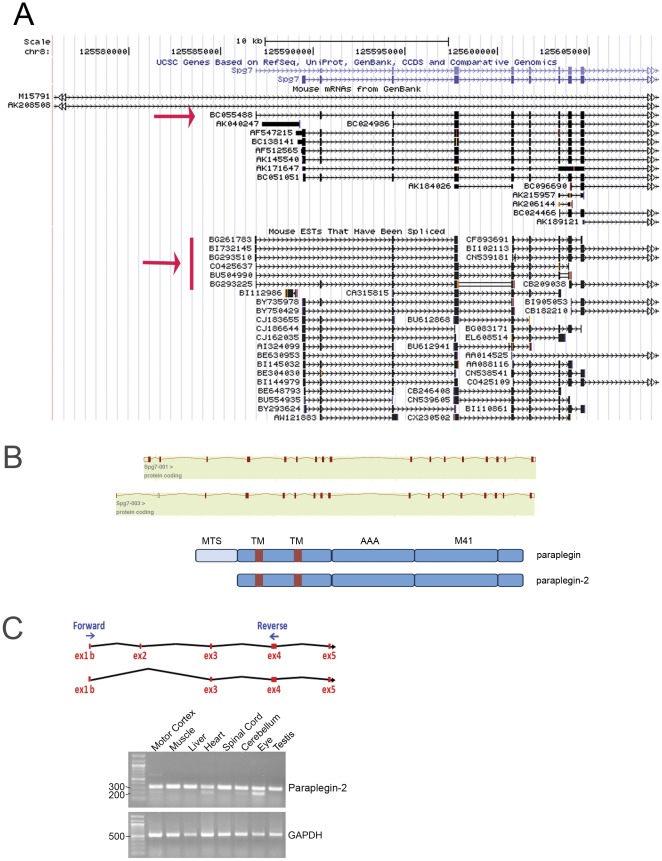
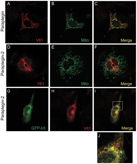
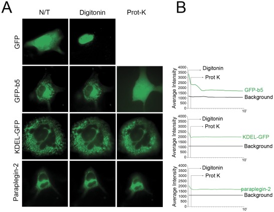
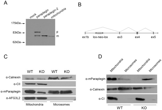
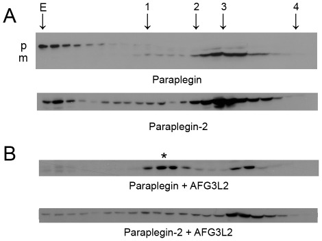
Principal findings: Here we show the existence in the mouse of a novel isoform of paraplegin, which we name paraplegin-2, encoded by alternative splicing of Spg7 through usage of an alternative first exon. Paraplegin-2 lacks the mitochondrial targeting sequence, and is identical to the mature mitochondrial protein. Remarkably, paraplegin-2 is targeted to the endoplasmic reticulum. We find that paraplegin-2 exposes the catalytic domains to the lumen of the endoplasmic reticulum. Moreover, endogenous paraplegin-2 accumulates in microsomal fractions prepared from mouse brain and retina. Finally, we show that the previously generated mouse model of Spg7-linked hereditary spastic paraplegia is an isoform-specific knock-out, in which mitochondrial paraplegin is specifically ablated, while expression of paraplegin-2 is retained.

Conclusions/significance: These data suggest a possible additional role of AAA proteases outside mitochondria and open the question of their implication in neurodegeneration.


Conflict of interest statement

Competing Interests: The authors have declared that no competing interests exist.

Figures

Figure 1
Figure 1. Alternative splicing isoforms of the murine Spg7 gene.
(A) Print-out from UCSC genome browser shows the existence of a complete cDNA (BC055488, red arrow) and six different ESTs (red bar) starting from an upstream alternative exon for the murine Spg7 gene. These ESTs present either ex1b-2 or ex1b-3 splicing and derives all from eye and retina libraries obtained at different developmental stages. (B) Schematic representation of the two alternative Spg7 transcripts obtained from the Ensembl database, and of the two respective predicted protein products. MTS, mitochondrial targeting sequence, TM, transmembrane domain, AAA, AAA domain, M41, M41 peptidase domain. (C) RT-PCR for the alternative Spg7 transcript on several mouse tissue cDNAs. On the top a scheme shows the position of the specific primers. The amplification products are detected as a 210 bp long or a 320 bp long DNA fragments, depending on the splicing pattern. Control amplification was performed using oligonucleotides specific for a housekeeping gene (GAPDH).
Figure 2
Figure 2. Subcellular localisation of paraplegin-2.
Immunofluorescence analysis of paraplegin and paraplegin-2 subcellular localisation in MEF cells after transfection of the respective cDNAs. While paraplegin decorates mitochondria (A–C), paraplegin-2 loses the mitochondrial localisation (D–F), but shows co-localisation with GFP-b5, a markers for the ER (G–I). Panel J shows an enlargement of the box in I. Paraplegin signal is detected using a specific antibody (V61). Mitochondria are labelled by overexpressing a mitochondrially targeted GFP.
Figure 3
Figure 3. Topology of paraplegin-2.
(A) FPP assay on MEFs transfected with different GFP fusion proteins. After permeabilization with digitonin (10 µM) for 1 min, all proteins, with the exception of a cytosolic GFP, retain their localisation in the ER. However, addition of 50 µg/ml proteinase K for 3 min disrupts the fluorescence pattern of cytosolic-exposed GFP-b5, but not that of KDEL-GFP and Paraplegin-2-GFP due to ER membrane protection. (B) Decrease in fluorescence of different GFP-fusion proteins was quantified in live imaging, using a cooled camera driven by METAMORPH software. Images were collected every 5 seconds for 10 minutes. Decrease in signal intensity after addition of proteinase K is visible, as expected, for GFP-b5, but not for KDEL-GFP, and paraplegin-2-GFP.
Figure 4
Figure 4. Paraplegin-2 is endogenously expressed in microsomal fractions.
(A) Western blot analysis of overexpressed paraplegin or paraplegin-2. Note that paraplegin-2 has the same electrophoretic motility as the mature form of paraplegin. Mitochondrial extracts are loaded as a control. p, precursor; m, mature form. (B) Scheme of the targeted allele in Spg7 −/− mice. (C) Western blot analysis of mitochondrial and microsomal fractions obtained from adult control and Spg7 −/− mouse brains. Enrichment of mitochondria and microsomes in the fraction were evaluated by using antibodies against 70 kDa subunit of Complex II and calnexin, respectively. A protein with the same molecular weight of paraplegin in the microsomal fractions is recognized by the specific paraplegin antibody. Note that 100 µg of microsomes were loaded versus 30 µg of mitochondria, and that blots were developed using ECL plus to reveal the bands in the microsomes. The paraplegin band in the mitochondrial fraction of Spg7−/− mice is likely a retrocontamination of mitochondria with microsomes. (D) Western blot analysis of mitochondrial and microsomal fractions obtained from retina of Spg7−/− and control mice using the specific paraplegin antibody. Pooled retinas from 15 Spg7 −/− mice and 12 control mice were used for this experiment. The enrichment of mitochondria and microsomes in the fractions was evaluated by antibodies against NDUFB6 (17 kDa subunit of Complex I) and calnexin, respectively.
Figure 5
Figure 5. Paraplegin-2 forms high-molecular weight complexes.
(A) Gel filtration analysis of paraplegin and paraplegin-2 high molecular weight complexes. Extracts obtained from COS7 cells transfected with paraplegin and paraplegin-2 cDNAs were fractionated by Superose 6 sizing chromatography. Eluate fractions were TCA precipitated and analysed by SDS-PAGE and immunoblotting using V61 α-paraplegin polyclonal antibodies. Overexpression of paraplegin or paraplegin-2 alone does not lead to the formation of a high molecular weight complex. Some aggregates are detected in fractions corresponding to the exclusion volume (E). In the case of paraplegin these aggregates are specific for the uncleaved precursor (p) that accumulates since cleavage of paraplegin depends on the m-AAA protease itself . (B) When AFG3L2 is co-expressed, the formation of a high molecular weight complex becomes evident for paraplegin (*), but not for paraplegin-2. Note that paraplegin is now completely cleaved. The following marker proteins were used for calibration: 1, Tyroglobulin (660 kDa); 2, Ferritin (440 kDa); 3, Alcohol Dehydrogenase (150 kDa); 4, Carbonic anhydrase (29 kDa), E indicates the exclusion volume of the column. p, precursor; m, mature form.

Similar articles

Cited by

References

    1. Harding AE. Hereditary spastic paraplegias. Semin Neurol. 1993;13:333–336. - PubMed
    1. Reid E, Rugarli EI. In: Valle, Beaudet, Vogelstein, Kinzler, Antonarakis, et al., editors. Scriver's Online Metabolic and Molecular Bases of Inherited Diseases; 2010.
    1. Casari G, De Fusco M, Ciarmatori S, Zeviani M, Mora M, et al. Spastic paraplegia and OXPHOS impairment caused by mutations in paraplegin, a nuclear-encoded mitochondrial metalloprotease. Cell. 1998;93:973–983. - PubMed
    1. Hansen JJ, Durr A, Cournu-Rebeix I, Georgopoulos C, Ang D, et al. Hereditary Spastic Paraplegia SPG13 Is Associated with a Mutation in the Gene Encoding the Mitochondrial Chaperonin Hsp60. Am J Hum Genet. 2002;70:1328–1332. - PMC - PubMed
    1. Arnoldi A, Tonelli A, Crippa F, Villani G, Pacelli C, et al. A clinical, genetic, and biochemical characterization of SPG7 mutations in a large cohort of patients with hereditary spastic paraplegia. Hum Mutat. 2008;29:522–531. - PubMed

Publication types

MeSH terms

Substances