. 2012 Mar 12;3(7):939-944.
doi: 10.1021/jz300150v.

Environment Polarity in Proteins Mapped Noninvasively by FTIR Spectroscopy

Affiliations

Environment Polarity in Proteins Mapped Noninvasively by FTIR Spectroscopy

Joshua Manor et al. J Phys Chem Lett. .

Abstract

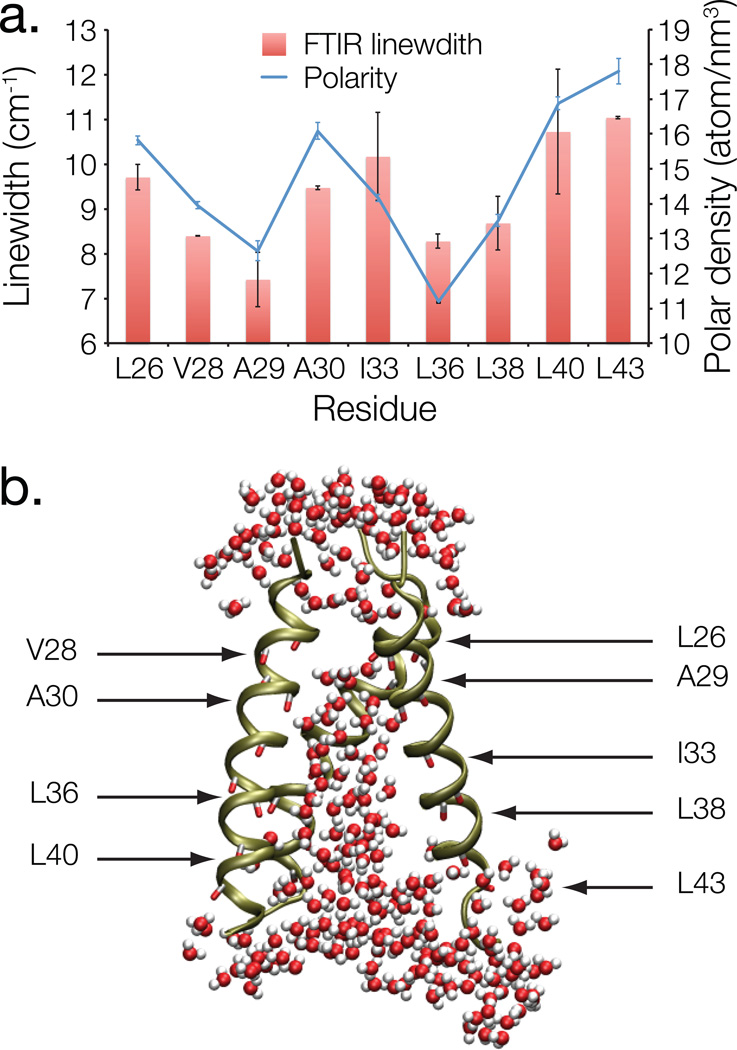
The polarity pattern of a macromolecule is of utmost importance to its structure and function. For example, one of the main driving forces for protein folding is the burial of hydrophobic residues. Yet polarity remains a difficult property to measure experimentally, due in part to its non-uniformity in the protein interior. Herein, we show that FTIR linewidth analysis of noninvasive 1-(13)C=(18)O labels can be used to obtain a reliable measure of the local polarity, even in a highly multi-phasic system, such as a membrane protein. We show that in the Influenza M2 H(+) channel, residues that line the pore are located in an environment that is as polar as fully solvated residues, while residues that face the lipid acyl chains are located in an apolar environment. Taken together, FTIR linewidth analysis is a powerful, yet chemically non-perturbing approach to examine one of the most important properties in proteins - polarity.


Figures

Figure 1
Figure 1
a. Sequence of the transmembrane peptides of the Influenza A M2 channel (S22-ÐL46) used in the current study. The red coloring points to the location of the 1-13C=18O labeled amino acids. b. Left panel: FTIR spectra of the M2 transmembrane peptide in hydrated lipid bilayers. The protein contains a single 1-13C=18O label at residue G34, which results in the isotope edited peak marked by an arrow. Right panel: Expansion of the spectra of the isotope edited peak depicting the two extreme possibilities for the baseline estimation. The linear baseline is depicted in red, while the constant baseline is shown in green. c. FTIR spectra (black) in the region of the 1-13C=18O isotope edited amide I mode of the M2 transmembrane peptides in hydrated lipid bilayers. Each panel presents the spectrum of a peptide that contains a single 1-13C=18O label at the indicated position. Peak fits (vertically shifted for visual clarity) according to the linear or constant baseline, alongside the resulting linewidth values are shown in red or green, respectively.
Figure 2
Figure 2
a. FTIR full width at half maximum linewidths (red columns) of nine 1-13C=18O amide groups in the influenza M2 H+ channel, obtained in hydrated lipid bilayers. The error bars are calculated as detailed in the experimental section. Environment polarity (blue line) was measured as the time-averaged density of polar atoms in a 12 Å radius sphere about the geometric center of the respective C=O group during the course of a 20 ns MD simulation. The error bars represent the standard deviation of the atom density with respect to time in 2 ns bins. b. Simulation snapshot of the M2 H+ channel in hydrated lipid bilayers. The protein is shown in ribbon form, alongside the isotopically labeled 1-13C=18O residues (indicated in licorice). Any water molecules that are within 4 Å of the protein are depicted in CPK representation. One of the helices was removed for visual clarity. The image was produced using VMD.

Similar articles

Cited by

References

    1. Langmuir I. Protein Monolayers. Cold Spring Harbor Symp Quant Biol. 1938;6:171–189.
    1. Tanford C. How Protein Chemists Learned About the Hydrophobic Factor. Protein Sci. 1997;6:1358–1366. - PMC - PubMed
    1. Isom DG, Castañeda CA, Cannon BR, García-Moreno EB. Large Shifts In pKa Values of Lysine Residues Buried Inside a Protein. Proc Natl Acad Sci U S A. 2011;108:5260–5265. - PMC - PubMed
    1. Pey AL, Rodriguez-Larrea D, Gavira JA, Garcia-Moreno B, Sanchez-Ruiz JM. Modulation of Buried Ionizable Groups In Proteins With Engineered Surface Charge. J Am Chem Soc. 2010;132:1218–1219. - PubMed
    1. Getahun Z, Huang C-Y, Wang T, De León B, DeGrado WF, Gai F. Using Nitrile-derivatized Amino Acids as Infrared Probes of Local Environment. J Am Chem Soc. 2003;125:405–411. - PubMed