

Early-stage visual processing abnormalities in high-functioning autism spectrum disorder (ASD)
- PMID: 22563527
- PMCID: PMC3342777
- DOI: 10.2478/v10134-010-0024-9
Early-stage visual processing abnormalities in high-functioning autism spectrum disorder (ASD)
Abstract
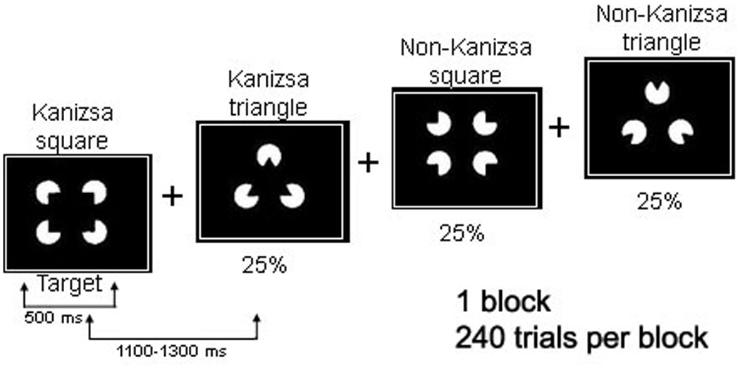
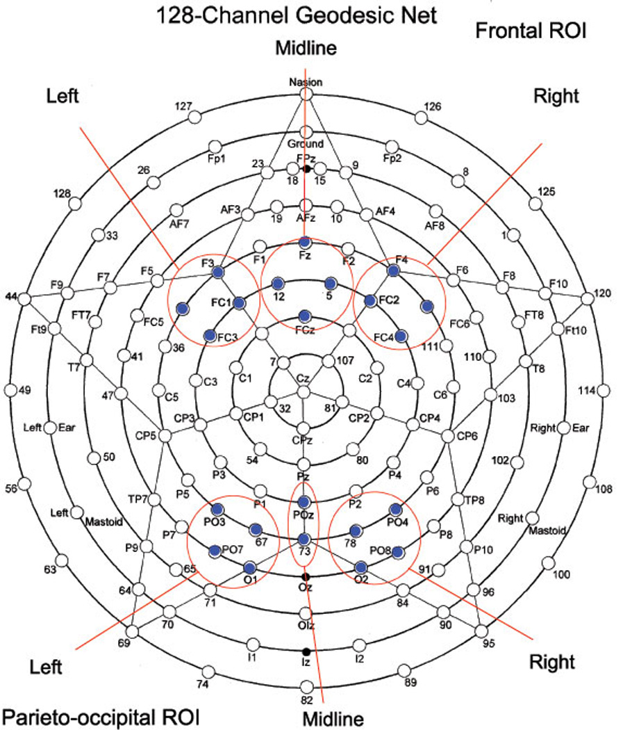
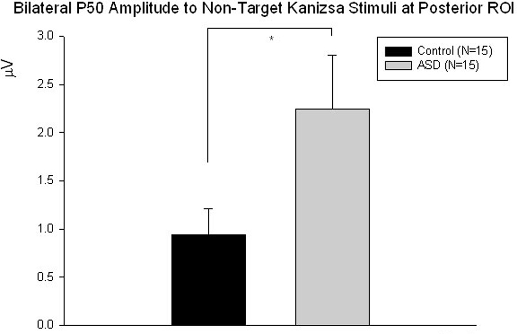
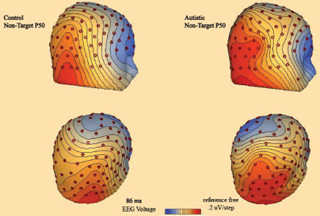
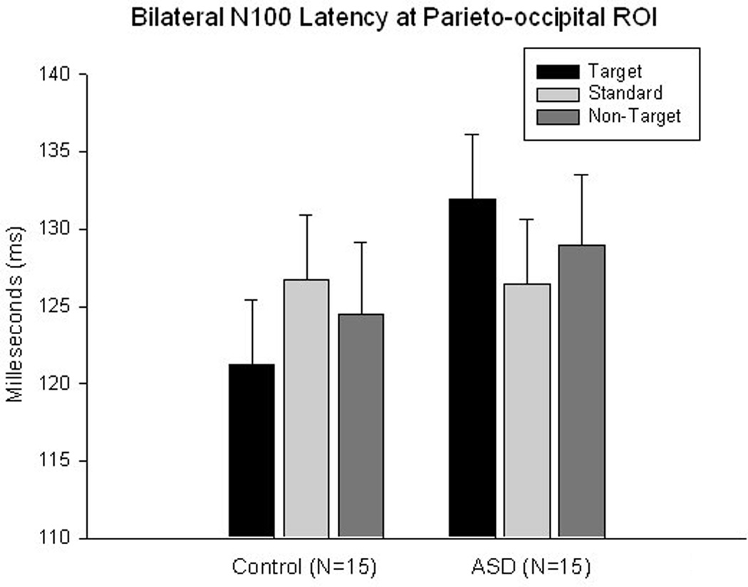
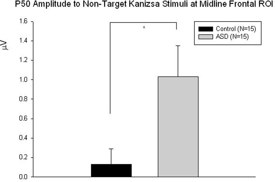
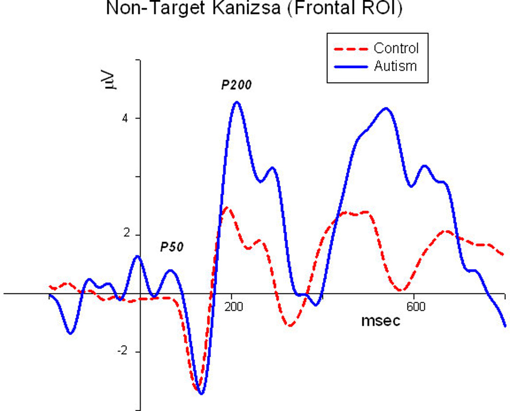
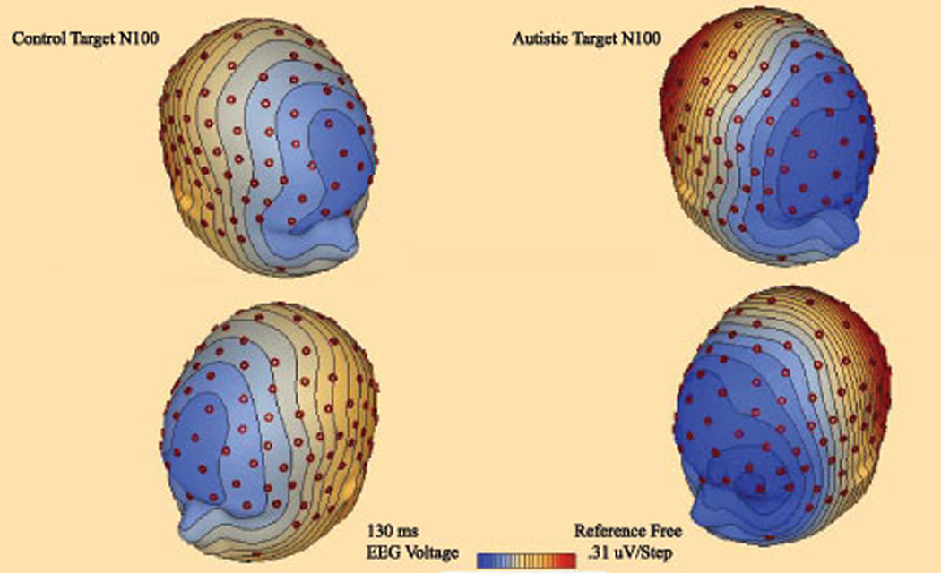
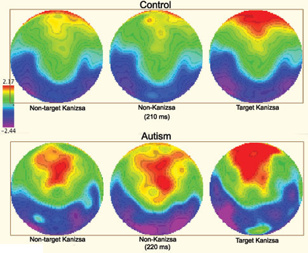
It has been reported that individuals with autism spectrum disorder (ASD) have abnormal responses to the sensory environment. For these individuals sensory overload can impair functioning, raise physiological stress, and adversely affect social interaction. Early-stage (i.e. within 200ms of stimulus onset) auditory processing abnormalities have been widely examined in ASD using event-related potentials (ERP), while ERP studies investigating early-stage visual processing in ASD are less frequent. We wanted to test the hypothesis of early-stage visual processing abnormalities in ASD by investigating ERPs elicited in a visual oddball task using illusory figures. Our results indicate that individuals with ASD have abnormally large cortical responses to task irrelevant stimuli over both parieto-occipital and frontal regions-of-interest (ROI) during early stages of visual processing compared to the control group. Furthermore, ASD patients showed signs of an overall disruption in stimulus discrimination, and had a significantly higher rate of motor response errors.
Figures
Similar articles
-
Atypical Processing of Novel Distracters in a Visual Oddball Task in Autism Spectrum Disorder.Behav Sci (Basel). 2017 Nov 16;7(4):79. doi: 10.3390/bs7040079. Behav Sci (Basel). 2017. PMID: 29144422 Free PMC article.
-
Event-related potential study of novelty processing abnormalities in autism.Appl Psychophysiol Biofeedback. 2009 Mar;34(1):37-51. doi: 10.1007/s10484-009-9074-5. Epub 2009 Feb 6. Appl Psychophysiol Biofeedback. 2009. PMID: 19199028
-
Repetitive Transcranial Magnetic Stimulation (rTMS) Modulates Event-Related Potential (ERP) Indices of Attention in Autism.Transl Neurosci. 2012 Jun 1;3(2):170-180. doi: 10.2478/s13380-012-0022-0. Transl Neurosci. 2012. PMID: 24683490 Free PMC article.
-
Translational use of event-related potentials to assess circuit integrity in ASD.Nat Rev Neurol. 2017 Mar;13(3):160-170. doi: 10.1038/nrneurol.2017.15. Epub 2017 Feb 17. Nat Rev Neurol. 2017. PMID: 28211449 Review.
-
Stimulus processing and error monitoring in more-able kindergarteners with autism spectrum disorder: a short review and a preliminary Event-Related Potentials study.Eur J Neurosci. 2018 Mar;47(6):556-567. doi: 10.1111/ejn.13580. Epub 2017 Jun 8. Eur J Neurosci. 2018. PMID: 28394438 Review.
Cited by
-
Perceptual category learning in autism spectrum disorder: Truth and consequences.Neurosci Biobehav Rev. 2020 Nov;118:689-703. doi: 10.1016/j.neubiorev.2020.08.016. Epub 2020 Sep 7. Neurosci Biobehav Rev. 2020. PMID: 32910926 Free PMC article. Review.
-
Subcortical evoked activity and motor enhancement in Parkinson's disease.Exp Neurol. 2016 Mar;277:19-26. doi: 10.1016/j.expneurol.2015.12.004. Epub 2015 Dec 11. Exp Neurol. 2016. PMID: 26687971 Free PMC article.
-
Development of social skills in children: neural and behavioral evidence for the elaboration of cognitive models.Front Neurosci. 2015 Sep 29;9:333. doi: 10.3389/fnins.2015.00333. eCollection 2015. Front Neurosci. 2015. PMID: 26483621 Free PMC article. Review.
-
Atypical Processing of Novel Distracters in a Visual Oddball Task in Autism Spectrum Disorder.Behav Sci (Basel). 2017 Nov 16;7(4):79. doi: 10.3390/bs7040079. Behav Sci (Basel). 2017. PMID: 29144422 Free PMC article.
-
Effects of weekly low-frequency rTMS on autonomic measures in children with autism spectrum disorder.Front Hum Neurosci. 2014 Oct 21;8:851. doi: 10.3389/fnhum.2014.00851. eCollection 2014. Front Hum Neurosci. 2014. PMID: 25374530 Free PMC article.
References
-
- American Psychiatric Association diagnostic and statistical manual of mental disorders (DSM-IV TR) 4th ed. Washington, D.C.: American Psychiatric Association; 2000.
-
- Charman T. Autism spectrum disorders. Psychiatry. 2008;7:331–334.
-
- Gomes E, Pedroso FS, Wagner MB. Auditory hypersensitivity in the autistic spectrum disorder. Pro Fono. 2008;20:279–284. - PubMed
-
- Khalfa S, Bruneau N, Rogé B, Georgieff N, Veuillet E, Adrien JL, et al. Increased perception of loudness in autism. Hear. Res. 2004;198:87–92. - PubMed
-
- Ratey JJ, Johnson C. The shadow syndromes. New York: Bantam Books; 1997.
Grants and funding
LinkOut - more resources
Full Text Sources