. 2012 Jun;38(5):223-32.
doi: 10.3109/01902148.2012.673049.

β-Nicotinamide adenine dinucleotide attenuates lipopolysaccharide-induced inflammatory effects in a murine model of acute lung injury

Affiliations

β-Nicotinamide adenine dinucleotide attenuates lipopolysaccharide-induced inflammatory effects in a murine model of acute lung injury

Nagavedi Siddaramappa Umapathy et al. Exp Lung Res. 2012 Jun.

Abstract

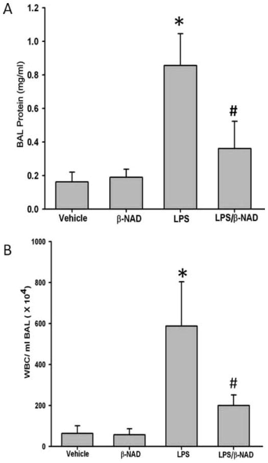
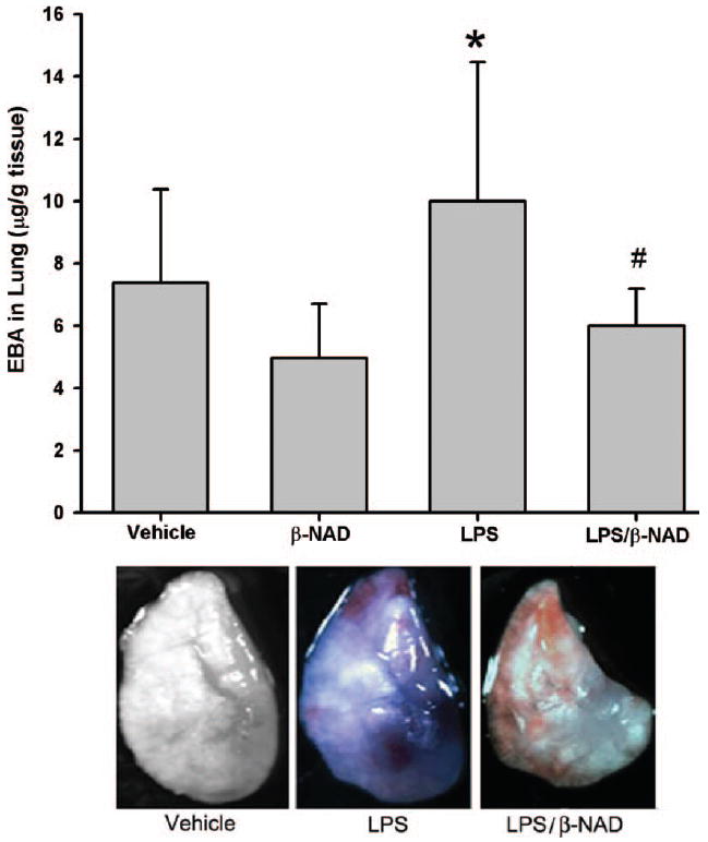
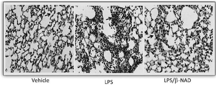
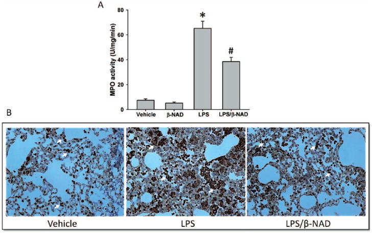
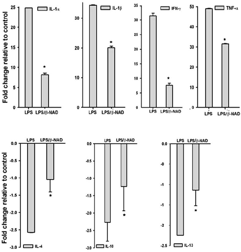
Acute lung injury (ALI) and acute respiratory distress syndrome (ARDS) occur in approximately 200,000 patients per year. Studies indicate that lung endothelium plays a significant role in ALI. The authors' recent in vitro studies demonstrate a novel mechanism of β-nicotinamide adenine dinucleotide (β-NAD)-induced protection against gram-positive (pneumolysin, PLY) and gram-negative (lipopolysaccharide, LPS) toxin-induced lung endothelial cell (EC) barrier dysfunction. The objective of the current study was to evaluate the protective effect of β-NAD against LPS-induced ALI in mice. C57BL/6J mice were randomly divided into 4 groups: vehicle, β-NAD, LPS, and LPS/β-NAD. After surgery, mice were allowed to recover for 24 hours. Evans blue dye-albumin (EBA) was given through the internal jugular vein 2 hours prior to the termination of the experiments. Upon sacrificing the animals, bronchoalveolar lavage fluid (BALF) was collected and the lungs were harvested. β-NAD treatment significantly attenuated the inflammatory response by means of reducing the accumulation of cells and protein in BALF, blunting the parenchymal neutrophil infiltration, and preventing capillary leak. In addition, the histological examination demonstrated decreased interstitial edema in the LPS/β-NAD specimens, as compared to the LPS-only specimens. The mRNA levels of the anti-inflammatory cytokines were up-regulated in the LPS group treated with β-NAD compared to the LPS-only-treated group. β-NAD treatment down-regulated the mRNA levels of the proinflammatory cytokines. These findings suggest that β-NAD could be investigated as a therapeutic option against bacterial toxin-induced lung inflammation and ALI in mice.


Conflict of interest statement

Declaration of interest: The authors report no conflicts of interest. The authors alone are responsible for the content and writing of the paper.

Figures

FIGURE 1
FIGURE 1
Bronchoalveolar lavage fluid (BALF) protein and cell counts. (A) BALF was collected at 24 hours after treatment, centrifuged, and protein was estimated in the clear supernatant using Bradford protein estimation kit. β-NAD treatment reduced total protein accumulation in the BALF of LPS-induced lung injury. The asterisk (*) indicates that a value significantly (P < .05) differs from the vehicle group and the number sign (#) indicates that a value significantly (P < .05) differs from LPS group (n = 4 for each group). (B) β-NAD reduces WBC accumulation in BALF of LPS-treated mice compared to untreated mice. The BALF was collected at 24 hours after treatment, centrifuged, and the cells were counted in hemocytometer. β-NAD reduced total WBCs in BALF. The asterisk (*) indicates that a value significantly (P < .001) differs from the vehicle group and the number sign (#) indicates that a value significantly (P < .05) differs from LPS group (n = 4 for each group). The error bars represent the standard error of the mean.
FIGURE 2
FIGURE 2
Evans blue dye–albumin (EBA) extravasations in BALF. (A) EBA was injected into the internal jugular vein 2 hours before the termination of the experiment. LPS challenge increased EBA leakage from the vascular space into surrounding lung tissue in the LPS group with notable attenuation in the LPS/β-NAD mice group. Both groups are compared to control and β-NAD-only–treated mice. The asterisk (*) indicates that a value significantly (P < .05) differs from the vehicle group and the number sign (#) indicates that a value significantly (P < .05) differs from LPS group (n = 4 for each group). The error bars represent the standard error of the mean. (B) β-NAD attenuated EBA leakage into the lung parenchyma on gross examination. Mice were grouped and the LPS group received LPS (0.9 mg/kg, i.t.) with PBS (i.v.), LPS/β-NAD group with LPS (0.9 mg/kg, i.t.) and β-NAD (5.46 mg/kg, i.v.), and control group with PBS (12 μL i.t. and 30 μL i.v.). EBA was injected into the right internal jugular vein 2 hours prior to termination of the experiment. The mice were sacrificed at 24 hours and immediately the lungs were flushed with EDTA, harvested, and photographed. Gross observation of the lung at 24 hours showed that the LPS/PBS lung exposure increased penetration of the EBA in the lung parenchyma, with minimal leakage in the LPS/β-NAD–treated specimen and none visible in vehicle.
FIGURE 3
FIGURE 3
Histopathology. β-NAD inhibits the inflammation in lungs of mice in LPS-induced ALI. Lungs perfused free of blood, were immersed in 4% buffered paraformaldehyde at 4°C for 18 hours prior to histological evaluation by hematoxylin and eosin (H&E) staining. H&E staining was done by deparafinizing and hydrating the slides to water. The slides were stained in Harris hematoxylin for 15 minutes and eosin for 30 seconds. The slides were dehydrated, cleared, and mounted with cytoseal. Histological analysis of the lung tissue obtained from the control mice exposed to PBS showed minimal infiltration of neutrophils. In contrast, mice exposed to LPS for 24 hours produced prominent neutrophil infiltration and that was attenuated in LPS/β-NAD simultaneously.
FIGURE 4
FIGURE 4
Myeloperoxidase (MPO) activity and staining. (A) Myeloperoxidaselevels in lung tissues of mice treated with or without β-NAD in LPS challenge. Neutrophil infiltration was analyzed by quantifying MPO levels in lungs tissues. β-NAD treatment significantly attenuates the MPO activity in lungs. (B) Myeloperoxidase staining was performed in lung tissues and the markedly increased infiltration of neutrophils (arrows) was observed in the lungs of mice from the LPS group, which was significantly attenuated by β-NAD treatment. The asterisk (*) indicates that a value significantly (P < .001) differs from the vehicle group and the number sign (#) indicates that a value significantly (P < .05) differs from LPS group (n = 4 for each group). The error bars represent the standard error of the mean.
FIGURE 5
FIGURE 5
Quantitative real-time PCR (qPCR). qPCR analysis of proinflammatory (upper panel) and anti-inflammatory (lower panel) cytokine gene expression from lungs of mice challenged with vehicle, LPS, and LPS/β-NAD. The bar represents the average fold change compared with vehicle and the expression levels were normalized to the value of housekeeping gene GAPDH mRNA. Data are shown as the mean ± SEM (n = 4 for each group). *P < .05 versus LPS group.

Similar articles

Cited by

References

    1. Abraham E. Neutrophils and acute lung injury. Crit Care Med. 2003;31(4 Suppl):S195–S199. - PubMed
    1. Razavi HM, Wang le F, Weicker S, Rohan M, Law C, McCormack DG, et al. Pulmonary neutrophil infiltration in murine sepsis: role of inducible nitric oxide synthase. Am J Respir Crit Care Med. 2004;170:227–233. - PubMed
    1. Wang le F, Patel M, Razavi HM, Weicker S, Joseph MG, McCormack DG, et al. Role of inducible nitric oxide synthase in pulmonary microvascular protein leak in murine sepsis. Am J Respir Crit Care Med. 2002;165:1634–1639. - PubMed
    1. Suda K, Tsuruta M, Eom J, Or C, Mui T, Jaw JE, et al. Acute lung injury induced cardiovascular dysfunction: effects of IL-6 and budesonide/formoterol. Am J Respir Cell Mol Biol. 2011 Jan 21;45:510–516. - PubMed
    1. Ware LB, Matthay MA. The acute respiratory distress syndrome. N Engl J Med. 2000;342:1334–1349. - PubMed

Publication types

MeSH terms