. 2012 May 24;55(10):4640-51.
doi: 10.1021/jm300460c. Epub 2012 May 16.

Discovery of the first irreversible small molecule inhibitors of the interaction between the vitamin D receptor and coactivators

Affiliations

Discovery of the first irreversible small molecule inhibitors of the interaction between the vitamin D receptor and coactivators

Premchendar Nandhikonda et al. J Med Chem. .

Abstract

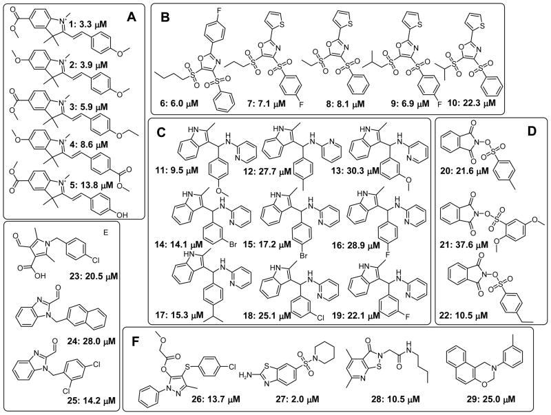
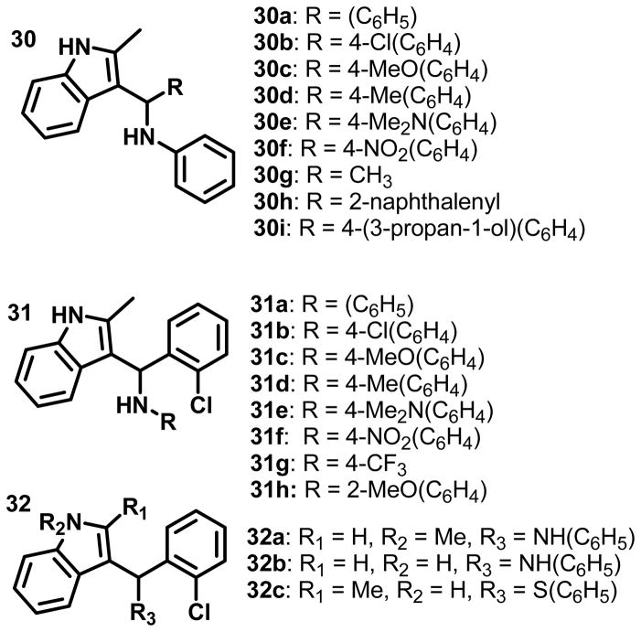
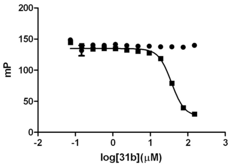
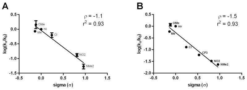
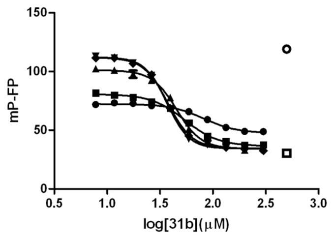
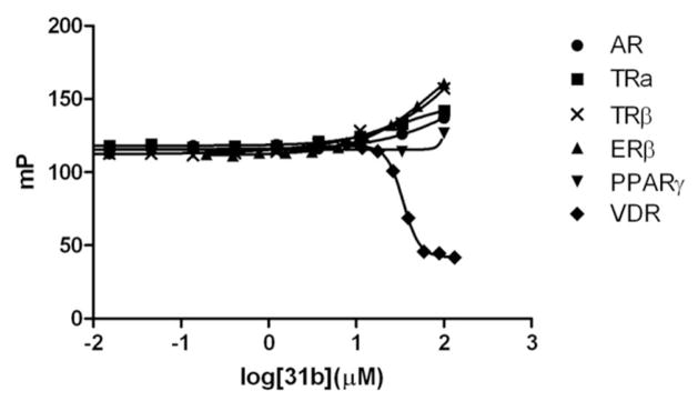
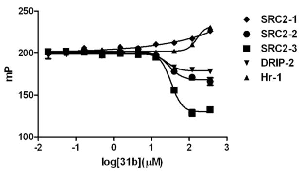
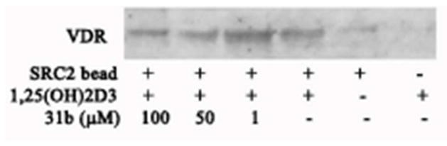
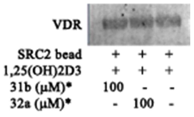
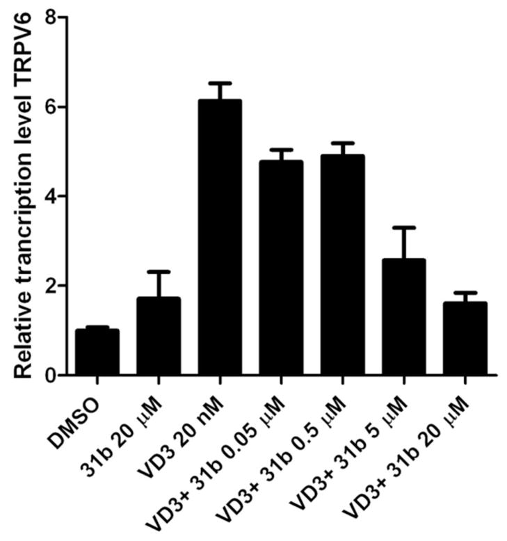
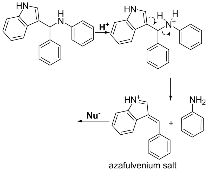
The vitamin D receptor (VDR) is a nuclear hormone receptor that regulates cell proliferation, cell differentiation, and calcium homeostasis. The receptor is activated by vitamin D analogues that induce the disruption of VDR-corepressor binding and promote VDR-coactivator interactions. The interactions between VDR and coregulators are essential for VDR-mediated transcription. Small molecule inhibition of VDR-coregulator binding represents an alternative method to the traditional ligand-based approach in order to modulate the expression of VDR target genes. A high throughput fluorescence polarization screen that quantifies the inhibition of binding between VDR and a fluorescently labeled steroid receptor coactivator 2 peptide was applied to discover the new small molecule VDR-coactivator inhibitors, 3-indolylmethanamines. Structure-activity relationship studies with 3-indolylmethanamine analogues were used to determine their mode of VDR-binding and to produce the first VDR-selective and irreversible VDR-coactivator inhibitors with the ability to regulate the transcription of the human VDR target gene TRPV6.


Figures

Figure 1
Figure 1
Hit structures from HTS for inhibitors of the interaction between VDR-LBD and fluorescently labeled peptide SRC2-3. Structures of validated hits are shown, grouped by chemotype, and annotated with IC50 values that were determined using a fluorescence polarization assay that employed VDR-LBD and fluorescently labeled peptide SRC2-3.
Figure 2
Figure 2
Structures of synthesized 3-indolyl-methanamines.
Figure 3
Figure 3
Identification of 31b’s reaction partner. ● Alexa Fluor-labeled SRC2-3 peptide (7 nM) was incubated for 3 hours with different concentration of 31b followed by the addition of VDR-LBD (1 μM) and LG190178 (5 μM). Fluorescence polarization was detected after 5 minutes. ■ VDR-LBD (1 μM) and LG190178 (5 μM) was incubated for 3 hours with different concentration of 31b followed by the addition of Alexa Fluor-labeled SRC2-3 peptide (7 nM). Fluorescence polarization was detected after 5 minutes.
Figure 4
Figure 4
Linear free energy relationship between VDR-SRC2-3 inhibition and the electronic nature of 3-indolyl-methanamines substitutents. A Series 30; B Series 31; log(kx/k0) values were calculated using rate constants given in Table 2. K0 being the non-substituted compounds 30a and 31a. σ-values were obtained from Ritchie et al. ρ-values represent the slopes of the linear regressions with the corresponding r2 values.
Figure 5
Figure 5
Influence of 2-mercaptoethanol for the inhibition of the VDR-SRC2-3 binding in the presence of 3-indolyl-methanamine, 31b. VDR-LBD (1 μM), LG190178 (5 μM), and Alexa Fluorlabeled SRC2-3 peptide (7 nM) were incubated in the presence of different concentrations of 31b and in the absence and presence of different concentrations of 2-mercaptoethanol. Interactions between VDR and SRC2-3 were determined by fluorescence polarization. ○ DMSO (negative control), □ 3- dibutylamino-1-(4-hexyl-phenyl)-propan-1-one (positive control), 2-mercaptoethanol concentrations (31b IC50 values): ● 100 mM (82.2 ± 7.3 μM), ■ 10 mM (54.5 ± 3.1 μM), ▲ 1 mM (45.4 ± 2.0 μM), ▼ 0.1 mM (37.5 ± 1.1 μM), ◆ 0.01 mM (36.8 ± 0.5 μM).
Figure 6
Figure 6
Nuclear receptor–coactivator binding studies in the presence of 3-indolyl-methanamine 31b using fluorescence polarization. FP was detected at an excitation/emission wavelength of 595/615 nm. The conditions for different NRs are as follows: AR: androgen receptor LBD (5 μM), Texas Redlabeled SRC2-3 (7 nM), and dihydrotestosterone (5 μM) were incubated with small molecule for 3h; TRα: thyroid receptor α LBD (2 μM), Texas Red-labeled SRC2-2 (7 nM), and triiodothyronine (1 μM) were incubated with small molecule for 3h; TRβ: thyroid receptor β LBD (0.8 μM), Texas Red-labeled SRC2-2 (7 nM), and triiodothyronine (1 μM) were incubated with small molecule for 3h; estrogen receptor β (3 μM), Texas Red-labeled SRC2-2 (5 nM), and estradiol (0.1 μM) were incubated with small molecule for 3h; peroxisome proliferator-activated receptor γ (5 μM), Texas Red-labeled DRIP2 (7 nM), and rosiglitazone (5 μM) were incubated with small molecule for 3h; VDR: vitamin D receptor LBD (1 μM), Texas Red-labeled SRC2-3 (7 nM), and LG190178 (5 μM) were incubated with small molecule for 3h.
Figure 7
Figure 7
VDR-coactivator interactions in the presence of 31b using FP. A VDR-LBD (1 μM), LG190178 (5 μM), and different Texas Red-labeled coregulator peptides (7 nM) were incubated for 3h in the presence of different concentrations of compound 31b.
Figure 8
Figure 8
Western Blot of in vitro binding reactions between SRC2 bearing all three NIDs and VDRLBD in the presence of 31b. Lane 1-3 different concentrations of 31b in the presence of VDR, SRC2 and 1,25(OH)2D3, lane 4 VDR, SRC2 and 1,25(OH)2D3, lane 5 no ligand (1,25(OH)2D3), no coregulator (SRC2).
Figure 9
Figure 9
Western Blot of in vitro binding reactions between SRC2 bearing all three NIDs and 3- indolylmethanamines. Lane 1 pre-incubation SRC2 with 31b, Lane 2 pre-incubation with 32a, and lane 3 pre-incubation with vehicle only. After incubation, beads were washed, treated with VDR and VDR–SRC2 interactions were determined by Western blot.
Figure 10
Figure 10
Modulation of expression of TRPV6 in the presence and absence of 1,25-(OH)2D3 and increasing concentrations of small molecule 31b. DU145 cells were cultured in six-well plates and treated with 1,25-(OH)2D3 (20 nM) and/or small molecule 31b. TRPV6 expression levels were determined by semi-quantitative RT-PCR and normalized to GAPDH transcript level and to DMSO control condition. The ΔΔCt method was used to measure the fold change in expression of genes. Standard deviations were calculated from three biological independent experiments performed in triplicates.
Scheme 1
Scheme 1
Proposed mechanism of action for 3-indolyl-methanamines.

Similar articles

Cited by

References

    1. Brumbaugh PF, Haussler MR. 1 Alpha,25-dihydroxycholecalciferol receptors in intestine. I. Association of 1 alpha,25-dihydroxycholecalciferol with intestinal mucosa chromatin. J Biol Chem. 1974;249:1251–1257. - PubMed
    1. Jurutka PW, Whitfield GK, Hsieh JC, Thompson PD, Haussler CA, Haussler MR. Molecular nature of the vitamin D receptor and its role in regulation of gene expression. Rev Endocr Metab Disord. 2001;2:203–216. - PubMed
    1. Feldman D, Pike JW, Glorieux FH. Vitamin D. 2. 1–2 Elsevier; Burlington: 2005.
    1. Yamada S, Shimizu M, Yamamoto K. Vitamin D receptor. Endocr Dev. 2003;6:50–63. - PubMed
    1. Toell A, Polly P, Carlberg C. All natural DR3-type vitamin D response elements show a similar functionality in vitro. Biochem J. 2000;352(Pt 2):301–309. - PMC - PubMed

Publication types

MeSH terms

LinkOut - more resources