. 2012 Jun 13;12(6):2773-7.
doi: 10.1021/nl204512x. Epub 2012 May 10.

Microcavity-integrated graphene photodetector

Affiliations
Free PMC article

Microcavity-integrated graphene photodetector

Marco Furchi et al. Nano Lett. .
Free PMC article

Abstract

There is an increasing interest in using graphene (1, 2) for optoelectronic applications. (3-19) However, because graphene is an inherently weak optical absorber (only ≈2.3% absorption), novel concepts need to be developed to increase the absorption and take full advantage of its unique optical properties. We demonstrate that by monolithically integrating graphene with a Fabry-Pérot microcavity, the optical absorption is 26-fold enhanced, reaching values >60%. We present a graphene-based microcavity photodetector with responsivity of 21 mA/W. Our approach can be applied to a variety of other graphene devices, such as electro-absorption modulators, variable optical attenuators, or light emitters, and provides a new route to graphene photonics with the potential for applications in communications, security, sensing and spectroscopy.


Figures
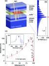
Figure 1
Figure 1
(a) Schematic drawing of a graphene microcavity photodetector. Distributed Bragg mirrors form a high-finesse optical cavity. The incident light is trapped in the cavity and passes multiple times through the graphene. The graphene sheet is shown in red, and the metal contacts are in yellow. (b) Electric field amplitude inside the cavity. (c) Calculated dependence of optical absorption in a single-layer graphene sheet on the reflectivity of the top mirror. The numbers next to the symbols indicate the number of SiO2/Si3N4 layer pairs that are necessary to achieve the respective reflectivity. Inset: Measured reflectivity of the AlAs/Al0.10Ga0.90As bottom mirror.
Figure 2
Figure 2
Reflectivity of the sample. The dip at 850 nm wavelength originates from absorption of the Fabry–Pérot microcavity mode.
Figure 3
Figure 3
(a) Photocurrent map taken at a bias voltage of VBias = 2 V between the source and drain electrodes. The gate electrode (substrate) remains unbiased. The dashed lines indicate the source and drain electrodes. The schematic above the photocurrent map illustrates the band diagram under this biasing condition. (b) Microscope image of a graphene photodetector and electrical setup for photocurrent measurements. The scale bar is 5 μm long. (c) Spectral response of the single-layer graphene device. The dashed lines show calculation results: reflection R (red), transmission T (green), and absorption A (blue). The solid lines are measurement results: reflection (red), photocurrent (blue). A strong and spectrally narrow photoresponse is observed at the cavity resonance (855 nm wavelength). Inset: Theoretical result for normal incidence light.
Figure 4
Figure 4
The meaning of the curves is the same as in Figure 3c, but the results are shown for a bilayer graphene device. A maximum responsivity of 21 mA/W is achieved. In addition, the spectral photoresponse of a conventional (without cavity) bilayer graphene detector is shown as solid red line. The response of the conventional device is approximately independent of wavelength, but more than an order of magnitude weaker than that of the microcavity device.

References

    1. Novoselov K. S.; Geim A. K.; Morozov S. V.; Jiang D.; Zhang Y.; Dubonos S. V.; Grigorieva I. V.; Firsov A. A. Science 2004, 306, 666–669. - PubMed
    1. Novoselov K. S.; Geim A. K.; Morozov S. V.; Jiang D.; Katsnelson M. I.; Grigorieva I. V.; Dubonos S. V.; Firsov A. A. Nature 2005, 438, 197–200. - PubMed
    1. Bonaccorso F.; Sun Z.; Hasan T.; Ferrari A. C. Nat. Photonics 2010, 4, 611–622.
    1. Blake P.; Brimicombe P. D.; Nair R. R.; Booth T. J.; Jiang D.; Schedin F.; Ponomarenko L. A.; Morozov S. V.; Gleeson H. F.; Hill E. W.; Geim A. K.; Novoselov K. S. Nano Lett. 2008, 8, 1704–1708. - PubMed
    1. Bae S.; Kim H.; Lee Y.; Xu X.; Park J.-S.; Zheng Y.; Balakrishnan J.; Lei T.; Kim H. R.; Song Y. I.; Kim Y.-J.; Kim K. S.; Özyilmaz B.; Ahn J.-H.; Hong B. H.; Iijima S. Nat. Nanotechnol. 2010, 5, 574–578. - PubMed

Publication types