. 2012 Jul 15;523(2):181-90.
doi: 10.1016/j.abb.2012.04.018. Epub 2012 Apr 30.

Differential roles of proteasome and immunoproteasome regulators Pa28αβ, Pa28γ and Pa200 in the degradation of oxidized proteins

Affiliations

Differential roles of proteasome and immunoproteasome regulators Pa28αβ, Pa28γ and Pa200 in the degradation of oxidized proteins

Andrew M Pickering et al. Arch Biochem Biophys. .

Abstract

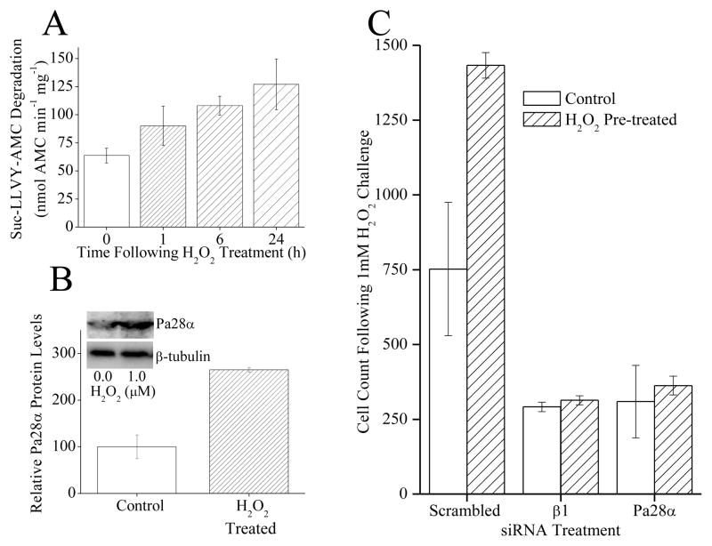
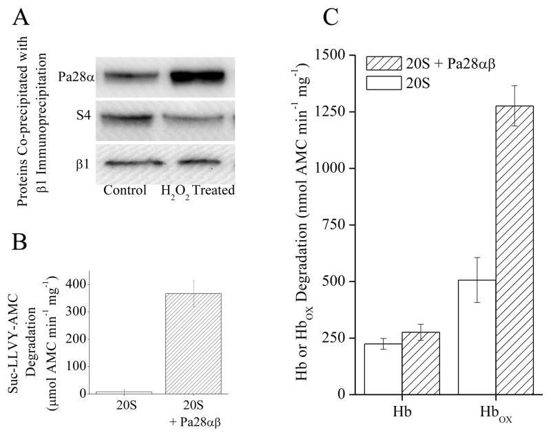
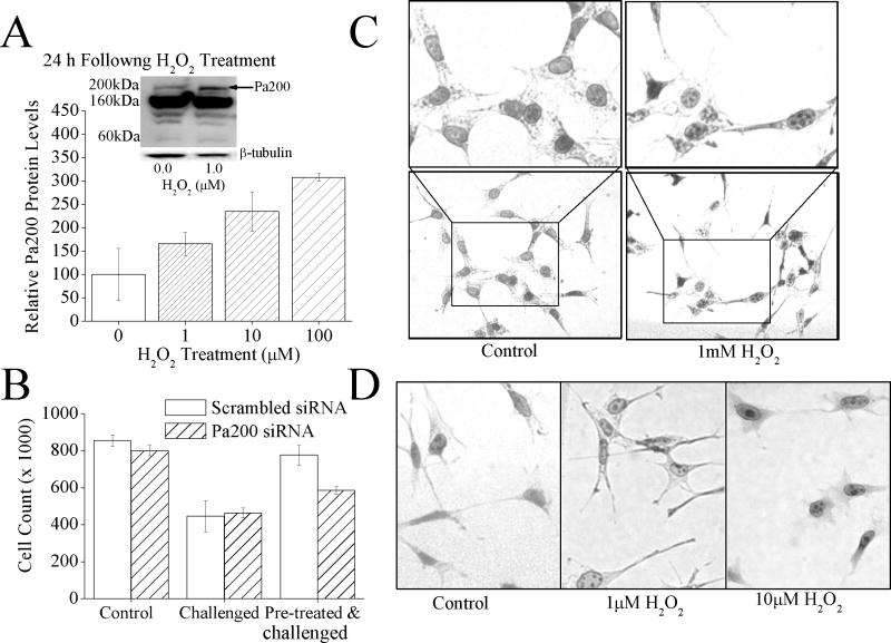
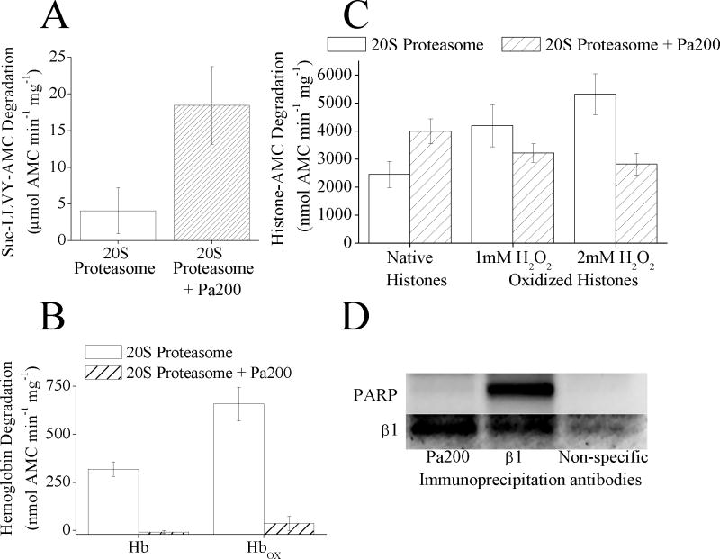
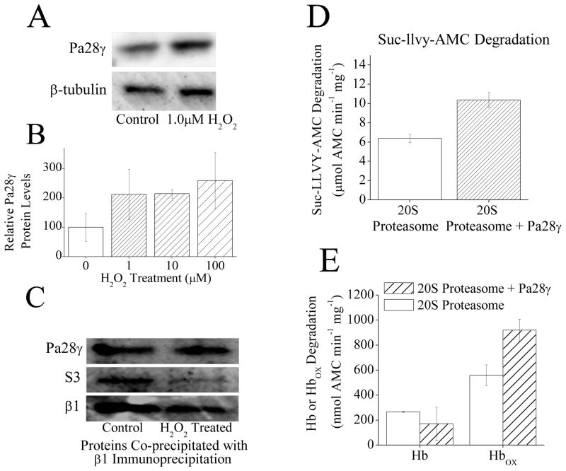
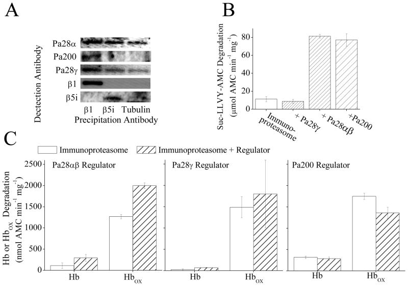
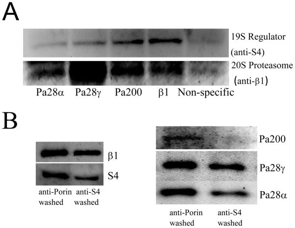
The response and functions of proteasome regulators Pa28αβ (or 11S), Pa28γ and Pa200 in oxidative-stress adaptation (also called hormesis) was studied in murine embryonic fibroblasts (MEFs), using a well-characterized model of cellular adaptation to low concentrations (1.0-10.0 μM) of hydrogen peroxide (H(2)O(2)), which alter gene expression profiles, increasing resistance to higher levels of oxidative-stress. Pa28αβ bound to 20S proteasomes immediately upon H(2)O(2)-treatment, whereas 26S proteasomes were disassembled at the same time. Over the next 24h, the levels of Pa28αβ, Pa28γ and Pa200 proteasome regulators increased during H(2)O(2)-adaptation, whereas the 19S regulator was unchanged. Purified Pa28αβ, and to a lesser extent Pa28γ, significantly increased the ability of purified 20S proteasome to selectively degrade oxidized proteins; Pa28αβ also increased the capacity of purified immunoproteasome to selectively degrade oxidized proteins but Pa28γ did not. Pa200 regulator actually decreased 20S proteasome and immunoproteasome's ability to degrade oxidized proteins but Pa200 and poly-ADP ribose polymerase may cooperate in enabling initiation of DNA repair. Our results indicate that cytoplasmic Pa28αβ and nuclear Pa28γ may both be important regulators of proteasome's ability to degrade oxidatively-damaged proteins, and induced-expression of both 20S proteasome and immunoproteasome, and their Pa28αβ and Pa28γ regulators are important for oxidative-stress adaptation.


Figures

Fig. 1
Fig. 1
(A) H2O2 exposure causes an increase in proteolytic capacity. MEF cells were pre-treated with 0, 1, 10 or 100μM H2O2. Then, 24 h later samples were harvested and lysed, and the capacity to degrade the short peptide Suc-LLVY-AMC was measured. Values are means ± SE where n = 3. (B) H2O2 exposure causes an increase in Pa28αβ protein levels. Samples were prepared as in panel A then run on Western blots which were screened with antibodies against either the α subunit of Pa28αβ or the loading control β-tubulin. (C) Blocking the induction of 20S proteasome β1 subunit, or Pa28ab proteasome regulator expression, with siRNAs, inhibited the H2O2 induced increase in oxidative stress tolerance, and blocked the increase in cell growth. siRNA knock-down with β1 siRNA, Pa28α siRNA, or scrambled siRNA was performed for 96 h. Cells were then pretreated with a mild dose of H2O2 then 24 h later challenged with a toxic dose of H2O2. Cell counts were taken 24 h later. Results are mean ± S.E. where n = 3.
Fig. 2
Fig. 2
(A), Following H2O2 treatment there is an increase in Pa28αβ association with 20S proteasome and a corresponding decrease in S4 (19S subunit) binding. MEF cells were treated with 1mM H2O2 for 1 h, after this immunoprecipitation of anti-β1 was performed. Samples were run on Western blots and screened for co-immunoprecipitation of anti-Pa28α, anti-S4, or anti-β1. (B) Pa28αβ increases the capacity of 20S proteasome to degrade the peptide substrate Suc-LLVY-AMC. 20S proteasome was incubated with a 4-fold molar excess of Pa28αβ for 30 minutes. Suc-LLVY-AMC was then added to samples and proteolytic capacity was measured. Values are means ± SE where n = 3. C. Pa28αβ increases the capacity of 20S proteasome to selectively degrade oxidized hemoglobin. 20S proteasome was incubated with a 4-fold molar excess of Pa28αβ for 30 minutes. Hb-AMC or HbOX-AMC was then added to samples and proteolytic capacity was measured. Values are means ± SE where n = 8.
Fig. 3
Fig. 3
(A) H2O2 exposure causes an increase in Pa200 protein levels. MEF cells were pretreated with 0, 1, 10, or 100μM H2O2, 24 h later samples were harvested and run on Western blots which were then screened with antibodies against either Pa200 or the loading control β-tubulin. Sample band intensity was measured and adjusted based on β-tubulin band intensity. Values are means ± SE where n = 3. The inset shows a representative blot. (B) By blocking the adaptive increase in Pa200, the H2O2 induced increase in oxidative stress tolerance is blunted. MEF cells treated with either Pa200 or (scrambled) siRNA for 24 h to block induction. Samples were then transiently adapted to oxidative stress by pre-treatment with 1μM of H2O2, for 1 h. Following a 24 h adaptation period, both adapted and non-adapted cells were challenged by incubation with a high dose of 100μM H2O2. Cell counts were then taken using a cell counter 24 h later. Results are means ± S.E. where n = 3. (C) Exposure to a toxic level of H2O2 causes the formation of Pa200 foci in the nucleus. MEF cells were exposed to 1.0mM H2O2 for 1hr. Immunocytochemistry was then performed and cells were stained with anti-Pa200. (D) Exposure to low, adaptive doses of H2O2 also results in the formation of nuclear Pa200 foci. Conditions were identical to those of panel C., except that 0, 1.0μM, or 10.0μM H2O2 exposures were used.
Fig. 4
Fig. 4
(A) Pa200 increases the capacity of 20S proteasome to degrade the peptide substrate Suc-LLVY-AMC. 20S proteasome was incubated with a 4 fold molar excess of Pa200 for 30 minutes. Suc-LLVY-AMC was then added to samples and proteolytic capacity measured over the next 4 h. Values are means ± SE where n = 3. (B) Pa200 reduces the capacity of 20S proteasome to degrade both oxidized and naive hemoglobin substrates. 20S Proteasome ± Pa200 was prepared as in panel A. Hb-AMC or HbOX-AMC was then added to samples and proteolytic capacity measured over the next 4 h. Values are means ± SE, where n = 8. (C) Pa200 enhances the capacity of proteasome to degrade normal histones but reduces proteasome’s capacity to degrade oxidized histones. 20S Proteasome ± Pa200 was prepared as in panel A and AMC labeled normal histones, or oxidized AMC labeled histones were used as substrates. Oxidized histones were prepared by treatment with either 1mM or 2mM H2O2 for 1 h. Proteolysis values are means ± SE, where n = 6. (D) Immunoprecipitation with either anti-Pa200 or anti-β1 both result in co- immunoprecipitation of 20S proteasome subunits, however only anti-β1 antibodies co-precipitate PARP. MEF cells were grown to 10% confluence, then immunoprecipitation with either anti-β1 antibody, anti-Pa200 antibody, or anti-porin antibody (as a non-specific binding control) was performed. Samples were run on Western blots and screened for co-immunoprecipitation of anti-β1 or anti-PARP.
Fig. 5
Fig. 5
(A) H2O2 exposure causes an increase in Pa28γ protein levels. MEF cells were pretreated with ± 1μM H2O2. Then, 24 h later samples were harvested and run on Western blots which were then screened with antibodies against either Pa200 or the loading control β-tubulin. (B) H2O2 exposure causes an increase in Pa28γ protein levels. MEF cells were prepared as in panel A in triplicate. Sample band intensity was then measured and adjusted based on β-tubulin band intensity. Values are means ± SE, where n = 3. (C) Following H2O2 treatment there is no change in Pa28γ association with 20S proteasome. MEF cells were exposed to 1mM H2O2 for 1 h, after which immunoprecipitation of anti-α5 was performed. Samples were run on Western blots and screened for co-immunoprecipitation of anti-Pa28γ, or anti-S3 (a subunit of the 19S regulator) as a measure of 26S proteasome disassembly. anti-α5 (not shown) was used as a loading control to confirm similar levels of 20S proteasome. The β1 blot was taken from a separate set of samples run under the same conditions. (D) Pa28γ slightly increases the capacity of 20S proteasome to degrade the peptide substrate Suc-LLVY-AMC. Purified 20S proteasome was incubated with a 4 fold molar excess of Pa28γ for 30 minutes. Suc-LLVY-AMC was then added to samples and proteolytic capacity measured. Values are means ± SE, where n = 3. (E) Pa28γ increases the capacity of 20S proteasome to degrade oxidized hemoglobin but not native hemoglobin. Purified 20S proteasome was incubated with a 4 fold molar excess of Pa28γ for 30 minutes. Hb-AMC or HbOX-AMC was then added to samples and proteolytic capacity measured. Values are means ± SE, where n = 8.
Fig. 6
Fig. 6
(A) Immunoprecipitation of MEF cell lysate with an antibody against the 20S proteasome subunit β1 causes co-precipitation of Pa28αβ, Pa200, and Pa28γ. However immunoprecipitation with an antibody against the immunoproteasome subunit β5i causes weak co-precipitation of Pa28αβ, and Pa28γ but not Pa200. Immunoprecipitation of MEF cells was performed using anti-β1, anti-β5i, or anti-β-tubulin (as a control for non-specific binding) antibodies. The Immunoprecipitate was analyzed by Western blotting and samples were screened for co-immunoprecipitation of anti-Pa28α, anti-Pa200, anti-Pa28γ, anti-β1, or anti-β5i. (B) Pa28αβ and Pa200 but not Pa28γ increase the capacity of immunoproteasome to degrade the peptide substrate Suc-LLVY-AMC. Immunoproteasome was incubated with a 4 fold molar excess of either Pa28γ Pa28αβ or Pa200 for 30 minutes. Suc-LLVY-AMC was then added to samples and proteolytic capacity measured. Values are means ± SE, where n = 3. (C) Binding of Pa28αβ (left panel) but not Pa28γ (center panel) or Pa200 (right panel) significantly increases the selective degradation of oxidized hemoglobin by immunoproteasome. Samples were prepared as in A, then AMC labeled hemoglobin or AMC labeled oxidized hemoglobin were added to samples as substrates. Values are means ± SE, where n = 8.
Fig. 7
Fig. 7
(A) Immunoprecipitation of MEF cell lysate with anti-Pa28α, anti-Pa28γ, anti-Pa200 or anti-β1 antibodies causes co-immunoprecipitation of both 20S and 26S proteasome subunits, however while anti-β1 and anti-Pa200 pull out a comparable amount of S4, anti-Pa28α and anti-Pa28γ antibodies are relatively poor at co- immunoprecipitation of S4. Immunoprecipitation protocols were as reported in the legends to Figs. 3 and 4. Non-specific binding was measured through immunoprecipitation of anti-porin. (B) Depletion of S4 through immunoprecipitation causes a small loss of Pa28α and Pa28γ but a very large loss of Pa200. MEF cell lysates were subjected to three cycles of immunoprecipitation with either anti-S4 or anti-porin. Non-immunoprecipitated supernatants were then taken and run on Western blots.

References

    1. Davies KJA. IUBMB Life. 2000;50:279–289. - PubMed
    1. Pickering AM, Koop AL, Teoh CY, Ermak G, Grune T, Davies KJA. Biochem J. 2010;432:585–594. - PMC - PubMed
    1. Grune T, Catalgol B, Licht A, Ermak G, Pickering AM, Ngo JK, Davies KJA. Free Radic Biol Med. 2011;51:1355–1364. - PMC - PubMed
    1. Davies KJA. Biochimie. 2001;83:301–310. - PubMed
    1. Reinheckel T, Sitte N, Ullrich O, Kuckelkorn U, Davies KJA, Grune T. Biochem J. 1998;335(Pt 3):637–642. - PMC - PubMed

Publication types

LinkOut - more resources