. 2012 Jul;97(7):2404-13.
doi: 10.1210/jc.2012-1274. Epub 2012 May 7.

Somatic GNAS mutation causes widespread and diffuse pituitary disease in acromegalic patients with McCune-Albright syndrome

Affiliations

Somatic GNAS mutation causes widespread and diffuse pituitary disease in acromegalic patients with McCune-Albright syndrome

Alexander O Vortmeyer et al. J Clin Endocrinol Metab. 2012 Jul.

Abstract

Context: McCune-Albright syndrome (MAS) is caused by sporadic mutations of the GNAS. Patients exhibit features of acromegaly. In most patients, GH-secreting pituitary adenomas have been held responsible for this presentation. However, surgical adenomectomy rarely eliminates excess GH production.

Objective: The aim of this study was to elucidate pituitary pathology in patients with MAS and to explain the basis of failure of adenomectomy to eliminate GH hypersecretion.

Design and setting: We conducted a case series at the National Institutes of Health.

Intervention(s): Interventions included medical therapy and transsphenoidal surgery.

Patients and main outcome measures: We studied clinical and imaging features and the histology and molecular features of the pituitary of four acromegalic MAS patients.

Results: We identified widespread and diffuse pituitary gland disease. The primary pathological changes were characterized by hyperplastic and neoplastic change, associated with overrepresentation of somatotroph cells in structurally intact tissue areas. Genetic analysis of multiple microdissected samples of any type of histological area consistently revealed identical GNAS mutations in individual patients. The only patient with remission after surgery received complete hypophysectomy in addition to removal of multiple GH-secreting tumors.

Conclusions: These findings indicate developmental effects of GNAS mutation on the entire anterior pituitary gland. The pituitary of individual cases contains a spectrum of changes with regions of normal appearing gland, hyperplasia, and areas of fully developed adenoma formation, as well as transitional stages between these entities. The primary change underlying acromegaly in MAS patients is somatotroph hyperplasia involving the entire pituitary gland, with or without development of somatotroph adenoma. Thus, successful clinical management, whether it is medical, surgical, or via irradiation, must target the entire pituitary, not just the adenomas evident on imaging.


Figures

Fig. 1.
Fig. 1.
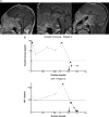
Patient 2, a 29-yr-old woman had acromegaly and extensive fibrous dysplasia of the frontal bones and the bones of the skull base with heterogeneous enhancement of the dysplastic bone (A, midline sagittal views; B, serial coronal views) on cranial and pituitary T1 MRI (A, left, midline sagittal view without enhancement; right, afterGd-DTPA administration). A and B, Dysplastic bone fills the sphenoid sinus region (*) and the entire anterior skull base (**); the arrows indicate the pituitary, and the arrowhead in right image indicates a nonenhancing microadenoma. B, Coronal views of pituitary MRI after contrast administration shows at least three nonenhancing microadenomas (arrows) within the anterior lobe on serial slices. C, Graph demonstrates the course of her GH and IGF-I levels over 3 yr. At surgery (arrow), three poorly defined microadenomas were removed with a portion of her surrounding anterior lobe, resulting in a transient drop in the levels of both hormones. Although IGF-I dropped into the normal range, the GH levels did not reach normal levels. After the presence of mammosomatotroph hyperplasia was evident, she was offered additional surgery for removal of the remainder of the gland, while the recently established surgical access through the dysplastic bone of the skull base was available, but she declined. Within 6 months, IGF-I was elevated again, coinciding with a rise in random GH levels. Follow-up MRI have been negative for additional tumor formation.
Fig. 2.
Fig. 2.
A, Contrast-enhanced sagittal MRI of patient 3, a 19-yr-old male with extensive, heterogeneously enhancing fibrous dysplasia of the bones of the skull base (left panel, *), hypogonadism, hypothyroidism, and acromegaly associated with a multinodular pituitary macroadenoma occupying his sella turcica and extending intracranially into the interpeduncular cistern and behind the clivus (arrow in center panel). The sella was surgically approached in stages; the first stage provided a midline transsphenoidal route to the sella (right panel). At the second stage, performed several days later, the approach was enlarged along the distal ventral edge, permitting exposure of a pituitary that contained multiple microadenomas and a moderately large macroadenoma. The tumors and the patient's pituitary were completely removed. B, Graph demonstrating the course of the patient's GH and IGF-I levels over 2 yr. He responded incompletely to octreotide (30 mg monthly) during the course of serial endocrine assessment (the black closed circles are values obtained while off medical therapy). Removal of his tumors and complete hypophysectomy (arrow) produced a prompt drop of his GH and IGF-I to 0.3 and 39 ng/ml, respectively. Regrettably, he had an intracranial hemorrhage 10 d after surgery from which he did not survive.
Fig. 3.
Fig. 3.
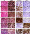
Structural and cytological changes in anterior pituitary gland tissue in patients with MAS; representative examples for different types of changes are illustrated by H&E stain, reticulin stain, and immunohistochemistry for GH and PRL, which were performed on adjacent sections. A, Mildly fibrotic, but otherwise regular anterior gland architecture with scattered somatotroph and lactotroph cells. B, Mildly fibrotic, but otherwise regular anterior gland architecture with predominantly somatotroph and only a few scattered lactotroph cells. C, Acidophilic hyperplasia, almost exclusively composed of somatotroph cells. D, Neoplasia with dominant somatotroph differentiation. E, Neoplasia with dominant lactrotroph differentiation. F, Neoplasia with predominantly mammosomatotroph differentiation.
Fig. 4.
Fig. 4.
A, In mammosomatotroph adenomas, mammosomatotroph differentiation was confirmed in tumor cells that demonstrated well-developed rough endoplasmic reticulum and characteristic granule extrusions (arrows). B and C, Double-immunolabeling of thin sections revealed positive signal for GH (large signal) in neuroendocrine granules of cells with somatotroph differentiation and positive granular signal for PRL (small signal) in cells with lactotroph differentiation. B, Mammosomatotroph cells, in contrast, revealed granules with immunoreactivity for both GH and PRL. C, Similarly, double-labeled mammosomatotroph cells were also detected in areas of acidophilic hyperplasia. The inset shows at higher magnification that positive signal for both GH and PRL are detected in neuroendocrine granules within the same cell. D, Rare scattered mammosomatotroph cells were detectable in regular anterior pituitary gland. A mammosomatotroph cell (right) is adjacent to a cell with exclusive immunoreactivity with anti-PRL (left).
Fig. 5.
Fig. 5.
A, Tissue microdissection; representative examples of microdissected normal tissue, hyperplastic tissue, and neoplastic tissue are shown. Before genetic analysis, different areas of normal, hyperplastic, and neoplastic anterior pituitary gland tissue were microdissected from H&E-stained slides; reticulin stains and immunohistochemical preparations from adjacent serial sections were used for identification of areas of interest. B, GNAS mutation analysis of pituitary gland tissue from three cases with MAS. DNA was extracted from microdissected tissue samples, amplified by PCR, and separately digested with NlaIII and PvuII, which can digest the PCR product in case the R201H mutation or the R201C of the GNAS gene is present, respectively. Case 1, Eleven foci of pituitary tissue were microdissected, mutation bands (arrow) are detected in different areas of hyperplasia and neoplasia, but not in three foci of normal-appearing pituitary gland (digestion with NlaIII). Case 2, Fifteen foci of pituitary tissue were microdissected; mutation bands (arrow) are detected in different areas of hyperplasia and neoplasia, but not in five different foci of normal-appearing pituitary gland (digestion with NlaIII). Case 3, Nine foci of pituitary gland tissue were microdissected; mutation bands (arrow) are detected in different areas of pituitary gland tissue with overrepresentation of somatotroph cells and areas of somatotroph hyperplasia (digestion with PvuII).

References

    1. de Sanctis C, Lala R, Matarazzo P, Balsamo A, Bergamaschi R, Cappa M, Cisternino M, de Sanctis V, Lucci M, Franzese A, Ghizzoni L, Pasquino AM, Segni M, Rigon F, Saggese G, Bertelloni S, Buzi F. 1999. McCune-Albright syndrome: a longitudinal clinical study of 32 patients. J Pediatr Endocrinol Metab 12:817–826 - PubMed
    1. Cuttler L, Jackson JA, Saeed uz-Zafar M, Levitsky LL, Mellinger RC, Frohman LA. 1989. Hypersecretion of growth hormone and prolactin in McCune-Albright syndrome. J Clin Endocrinol Metab 68:1148–1154 - PubMed
    1. Pun KK, Chan G, Kung A, Lam K, Chan FL, Wang C. 1989. McCune-Albright syndrome with acromegaly. Horm Metab Res 21:527–528 - PubMed
    1. Chanson P, Dib A, Visot A, Derome PJ. 1994. McCune-Albright syndrome and acromegaly: clinical studies and responses to treatment in five cases. Eur J Endocrinol 131:229–234 - PubMed
    1. Bhansali A, Sharma BS, Sreenivasulu P, Singh P, Vashisth RK, Dash RJ. 2003. Acromegaly with fibrous dysplasia: McCune-Albright Syndrome—clinical studies in 3 cases and brief review of literature. Endocr J 50:793–799 - PubMed

MeSH terms