. 2012 Jun;122(6):2104-13.
doi: 10.1172/JCI61228. Epub 2012 May 8.

Protein disulfide isomerase inhibitors constitute a new class of antithrombotic agents

Affiliations

Protein disulfide isomerase inhibitors constitute a new class of antithrombotic agents

Reema Jasuja et al. J Clin Invest. 2012 Jun.

Abstract

Thrombosis, or blood clot formation, and its sequelae remain a leading cause of morbidity and mortality, and recurrent thrombosis is common despite current optimal therapy. Protein disulfide isomerase (PDI) is an oxidoreductase that has recently been shown to participate in thrombus formation. While currently available antithrombotic agents inhibit either platelet aggregation or fibrin generation, inhibition of secreted PDI blocks the earliest stages of thrombus formation, suppressing both pathways. Here, we explored extracellular PDI as an alternative target of antithrombotic therapy. A high-throughput screen identified quercetin-3-rutinoside as an inhibitor of PDI reductase activity in vitro. Inhibition of PDI was selective, as quercetin-3-rutinoside failed to inhibit the reductase activity of several other thiol isomerases found in the vasculature. Cellular assays showed that quercetin-3-rutinoside inhibited aggregation of human and mouse platelets and endothelial cell-mediated fibrin generation in human endothelial cells. Using intravital microscopy in mice, we demonstrated that quercetin-3-rutinoside blocks thrombus formation in vivo by inhibiting PDI. Infusion of recombinant PDI reversed the antithrombotic effect of quercetin-3-rutinoside. Thus, PDI is a viable target for small molecule inhibition of thrombus formation, and its inhibition may prove to be a useful adjunct in refractory thrombotic diseases that are not controlled with conventional antithrombotic agents.


Figures

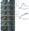
Figure 1
Figure 1. Identification of quercetin-3-rutinoside as a PDI inhibitor.
(A) Data from representative 384-well plates, including samples with no PDI (green); 3 mM bacitracin, a nonspecific inhibitor of the PDI family of oxidoreductases (blue); or test compounds (red). Duplicate readings are plotted, and an arrow indicates the inhibitory compound identified as quercetin-3-rutinoside. Each symbol represents an individual reading. (B) Effect of the indicated concentrations of quercetin-3-rutinoside on PDI activity, determined using the insulin reduction assay (mean ± SD). (C) Sensorgram of binding of quercetin-3-rutinoside to recombinant PDI. The thin arrow indicates the time of injection of quercetin-3-rutinoside onto the PDI-coated sensor chip, and the thick arrow indicates the time of injection of regeneration buffer. Black lines represent the fitted curves for duplicates of the varying concentrations of quercetin-3-rutinoside analyte at 0, 0.39, 0.78, 1.56, 3.13, 6.25, 12.5, 25, 50, and 100 μM.
Figure 2
Figure 2. Structure-activity relationship of the flavonols and their potency (IC50) of PDI inhibition.
Numbers in structure correspond with those in the column headings.
Figure 3
Figure 3. Quercetin-3-rutinoside selectively inhibits PDI.
(A) Effect of quercetin-3-rutinoside at 0.3 μM (red), 3 μM (blue), and 30 μM (green) on PDI, thioredoxin (TRX), ERp5, ERp72, and ERp57 activity, as measured in the insulin reduction assay. Data show the mean and SD of 3 independent determinations. (B) Insulin reductase assay in the absence (black) or presence (red) of 30 μM quercetin-3-rutinoside in addition to thioredoxin, thioredoxin reductase, and NADPH as reducing equivalent.
Figure 4
Figure 4. Quercetin-3-rutinoside inhibits platelet aggregation.
(A) Washed human platelets (2 × 10⁸ platelets/ml) were incubated with vehicle alone (black), 30 μM quercetin-3-rutinoside (blue), 45 μM quercetin-3-rutinoside (red), or anti-PDI antibody, RL90 (green), for 15 minutes and subsequently stimulated with 50 μM PAR4 peptide AYPGKF. (B) Washed human platelets (2 × 10⁸ platelets/ml) were incubated with either vehicle (black) or 60 μM quercetin-3-rutinoside (red) for 15 minutes or incubated with vehicle followed by a wash (dark blue) or 60 μM quercetin-3-rutinoside followed by a wash (light blue) and subsequent stimulation with 50 μM PAR4 peptide AYPGKF. (C) Quercetin-3-rutinoside was infused into mice intravenously at 0.5 mg/kg. Five minutes after infusion, blood was obtained by cardiac puncture, and platelet-rich plasma was isolated. Platelet-rich plasma from quercetin-3-rutinoside–treated mice (red) or vehicle control (black) was stimulated with 200 μM AYPGKF.
Figure 5
Figure 5. Quercetin-3-rutinoside inhibits fibrin generation in vitro.
(A and B) Representative images of fixed and immunostained HUVECs that have been activated by laser injury in the presence of plasma and calcium (B) with or (A) without quercetin-3-rutinoside (10 μM). The cells were fixed after laser activation and stained for fibrin (red), FITC-phalloidin (green), and DAPI (blue). (C) Quantification of fibrin signal detected on cultured endothelial cells expressed as the percentage inhibition of fibrin after laser activation (mean ± SD). **P < 0.01, ***P < 0.001. Original magnification, ×60. Scale bars: 10 μm.
Figure 6
Figure 6. Quercetin-3-rutinoside inhibits thrombus formation and fibrin generation in vivo.
Platelet-specific anti-CD42b antibody conjugated to Dylight 649 (0.1 μg/g body weight) and fibrin-specific mouse anti-human fibrin II β-chain monoclonal antibody conjugated to Alexa Fluor 488 (0.5 μg/g body weight) were infused into the mice. At varying doses, quercetin-3-rutinoside was subsequently infused intravenously 5 minutes prior to the initial laser injury. Representative binarized images of the appearance of fluorescence signals associated with fibrin (green) and platelets (red) over the 180 seconds after laser-induced vessel wall injury in wild-type mice are shown in AD, with mice infused with (A) vehicle only; (B) quercetin-3-rutinoside at 0.1 mg/kg body weight; (C) quercetin-3-rutinoside at 0.3 mg/kg body weight; and (D) quercetin-3-rutinoside at 0.5 mg/kg body weight shown. (E) Median integrated platelet fluorescence intensity and (F) median integrated fibrin fluorescence intensity at the injury site are plotted versus time, with mice infused with vehicle only (black); quercetin-3-rutinoside at 0.1 mg/kg body weight (green); quercetin-3-rutinoside at 0.3 mg/kg body weight (blue); and quercetin-3-rutinoside at 0.5 mg/kg body weight shown (red). Data are from 30 thrombi in 3 mice for each condition. RFU, relative fluorescence units. (G) Quercetin-3-rutinoside at 0.5 mg/kg was infused 5 minutes prior to FeCl3 injury of cremaster arterioles. Data points represent time to occlusion, as determined by cessation of flow in control mice infused with vehicle or mice infused with quercetin-3-rutinoside as indicated. Horizontal bars denotes median values. Original magnification, ×60. Scale bars: 10 μm.
Figure 7
Figure 7. Oral quercetin-3-rutinoside inhibits thrombus formation and fibrin generation in vivo.
Platelet-specific anti-CD42b antibody conjugated to Dylight 649 (0.1 μg/g body weight) and fibrin-specific mouse anti-human fibrin II β-chain monoclonal antibody conjugated to Alexa Fluor 488 (0.5 μg/g body weight) were infused into the mice. (A) Median integrated platelet fluorescence and (B) fibrin fluorescence at the injury site after oral gavage with quercetin-3-rutinoside 90 minutes prior to injury are plotted versus time, with mice gavaged with vehicle only (black); with 5 mg/kg quercetin-3-rutinoside (red); with 10 mg/kg quercetin-3-rutinoside (blue); with 20 mg/kg quercetin-3-rutinoside (green); and with 50 mg/kg quercetin-3-rutinoside (purple). Data are from 30 thrombi in 3 mice for each condition.
Figure 8
Figure 8. The quercetin analog diosmetin does not affect thrombus formation and fibrin generation in vivo.
Platelet-specific anti-CD42b antibody conjugated to Dylight 649 (0.1 μg/g body weight) and fibrin-specific mouse anti-human fibrin II β-chain monoclonal antibody conjugated to Alexa Fluor 488 (0.5 μg/g body weight) were infused into the mice. Diosmetin at 10 mg/kg body weight or vehicle control was subsequently infused intravenously immediately prior to the initial laser injury. Representative binarized images of the appearance of fluorescence signals associated with fibrin (green) and platelets (red) over 180 seconds after laser-induced vessel wall injury in a wild-type mouse are shown in A and B, with mice infused with (A) vehicle only or (B) diosmetin at 10 mg/kg body weight. (C) Median integrated platelet fluorescence intensity and (D) median integrated fibrin fluorescence intensity at the injury site are plotted versus time, with mice infused with vehicle only (black) or 10 mg/kg diosmetin (blue). Data are from 30 thrombi in 3 mice for each condition. Original magnification, ×60. Scale bars: 10 μm.
Figure 9
Figure 9. Exogenous PDI reverses quercetin-3-rutinoside inhibition of thrombus formation and fibrin generation in vivo.
Dylight 649–conjugated anti-CD42b antibody and Alexa Fluor 488–conjugated fibrin-specific antibody were infused into mice. Mice were subsequently infused with (A and D) vehicle alone or (B and C) 0.25 mg/kg quercetin-3-rutinoside. After 6 to 12 initial thrombi, mice were infused with either (A and B) vehicle or (C and D) PDI at 200 μg per mouse to record additional thrombi. Representative binarized images of the appearance of fluorescence signals associated with fibrin (green) and platelets (red) over 180 seconds after laser-induced vessel wall injury. (E) Median integrated platelet fluorescence and (F) median integrated fibrin fluorescence at the injury site in mice infused with vehicle only (black), 0.25 mg/kg quercetin-3-rutinoside followed by vehicle (green), vehicle followed by 200 μg recombinant PDI (red), or 0.25 mg/kg quercetin-3-rutinoside followed by 200 μg recombinant PDI (blue). Data are from 30 thrombi in 3 mice for each condition. Original magnification, ×60. Scale bars: 10 μm.

Similar articles

Cited by

References

    1. Hatahet F, Ruddock LW. Protein disulfide isomerase: a critical evaluation of its function in disulfide bond formation. Antioxid Redox Signal. 2009;11(11):2807–2850. doi: 10.1089/ars.2009.2466. - DOI - PubMed
    1. Manickam N, Sun X, Li M, Gazitt Y, Essex DW. Protein disulphide isomerase in platelet function. Br J Haematol. 2008;140(2):223–229. - PubMed
    1. Hotchkiss KA, Matthias LJ, Hogg PJ. Exposure of the cryptic Arg-Gly-Asp sequence in thrombospondin-1 by protein disulfide isomerase. Biochim Biophys Acta. 1998;1388(2):478–488. doi: 10.1016/S0167-4838(98)00211-8. - DOI - PubMed
    1. Essex DW, Li M. Protein disulphide isomerase mediates platelet aggregation and secretion. Br J Haematol. 1999;104(3):448–454. doi: 10.1046/j.1365-2141.1999.01197.x. - DOI - PubMed
    1. Burgess JK, et al. Physical proximity and functional association of glycoprotein 1balpha and protein-disulfide isomerase on the platelet plasma membrane. J Biol Chem. 2000;275(13):9758–9766. doi: 10.1074/jbc.275.13.9758. - DOI - PubMed

Publication types

MeSH terms