Clinical Trial
. 2012 Jun;122(6):2221-33.
doi: 10.1172/JCI59535. Epub 2012 May 8.

IL-15 protects NKT cells from inhibition by tumor-associated macrophages and enhances antimetastatic activity

Affiliations
Clinical Trial

IL-15 protects NKT cells from inhibition by tumor-associated macrophages and enhances antimetastatic activity

Daofeng Liu et al. J Clin Invest. 2012 Jun.

Abstract

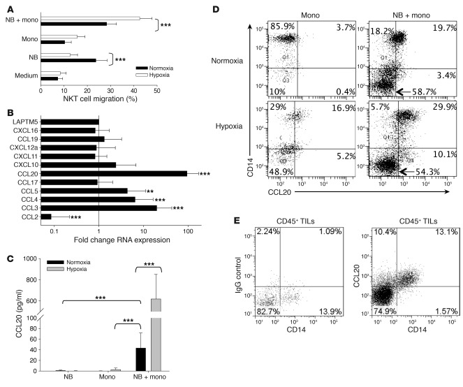
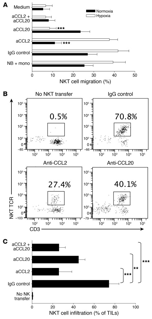
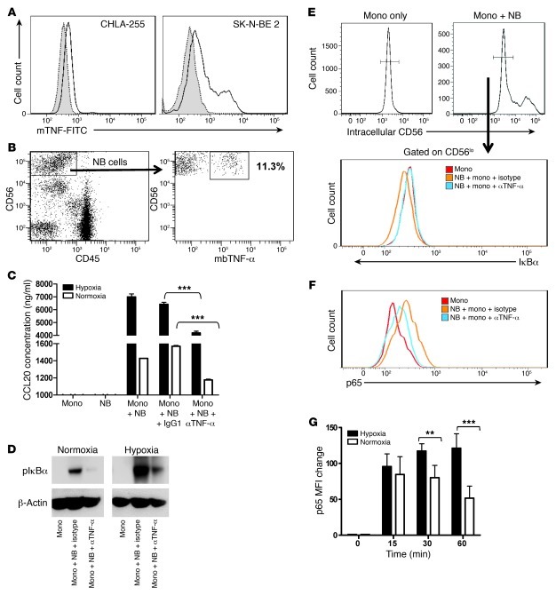
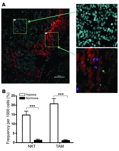
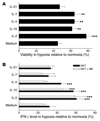
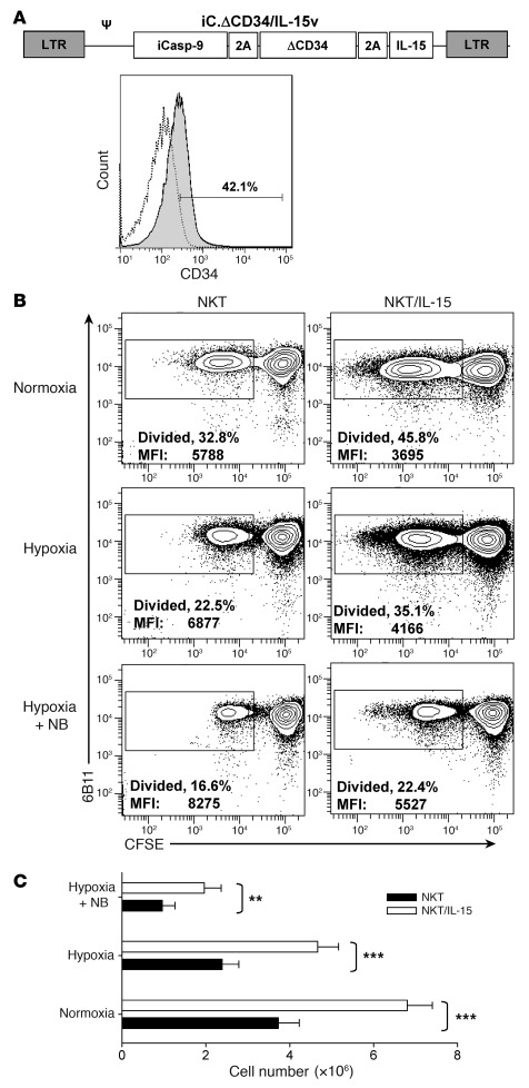
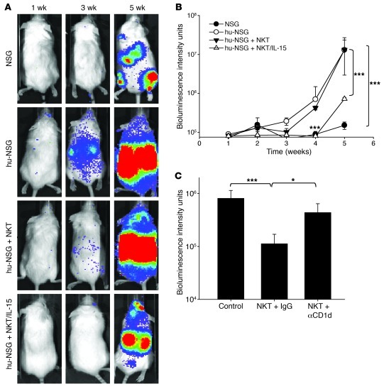
Vα24-invariant NKT cells inhibit tumor growth by targeting tumor-associated macrophages (TAMs). Tumor progression therefore requires that TAMs evade NKT cell activity through yet-unknown mechanisms. Here we report that a subset of cells in neuroblastoma (NB) cell lines and primary tumors expresses membrane-bound TNF-α (mbTNF-α). These proinflammatory tumor cells induced production of the chemokine CCL20 from TAMs via activation of the NF-κB signaling pathway, an effect that was amplified in hypoxia. Flow cytometry analyses of human primary NB tumors revealed selective accumulation of CCL20 in TAMs. Neutralization of the chemokine inhibited in vitro migration of NKT cells toward tumor-conditioned hypoxic monocytes and localization of NKT cells to NB grafts in mice. We also found that hypoxia impaired NKT cell viability and function. Thus, CCL20-producing TAMs served as a hypoxic trap for tumor-infiltrating NKT cells. IL-15 protected antigen-activated NKT cells from hypoxia, and transgenic expression of IL-15 in adoptively transferred NKT cells dramatically enhanced their antimetastatic activity in mice. Thus, tumor-induced chemokine production in hypoxic TAMs and consequent chemoattraction and inhibition of NKT cells represents a mechanism of immune escape that can be reversed by adoptive immunotherapy with IL-15-transduced NKT cells.


Figures

Figure 1
Figure 1. Contact with NB cells and hypoxia synergistically induce CCL20 in human monocytes.
(A) Primary monocytes were cocultured with CHLA-255 NB cells (1:1 ratio) for 48 hours in normoxic (20% O2) or hypoxic (1% O2) conditions, and supernatants were placed in bottom chambers of dual-chambers plates with 5-μM pore membranes with or without addition of the indicated neutralizing antibodies or their isotype control. NKT cells were placed in the upper chambers and allowed to migrate for 3 hours, and the rate of NKT cell migration was quantified by FACS. Results are mean ± SD from 3 experiments in triplicate. (B) Monocytes were cocultured with or without CHLA-255 NB cells for 36 hours in normoxic or hypoxic conditions followed by mRNA isolation and quantitative real-time PCR analysis of 11 chemokine genes known to attract human NKT cells. Data are from a representative of 3 experiments in triplicate. (C) Monocytes and CHLA-255 NB cells were cultured alone or combined in hypoxic or normoxic conditions for 48 hours. CCL20 concentration was quantified in the supernatants using ELISA. Data are mean ± SD from experiments with monocytes from 6 donors in duplicate. (D) Cells were cultured as in C and analyzed for intracellular CCL20 accumulation in CD14+ monocytes and CD14 NB cells. Regions were set using corresponding isotype controls. Data are from a representative of 3 experiments in duplicate. (E) Tumor-infiltrating leukocytes (TILs) were isolated from a cell suspension of freshly resected primary NB by gradient centrifugation and cultured with GolgiStop for 4 hours followed by FACS. After gating on CD45+ events, CCL20 accumulation was examined in CD14+ TAMs and compared with the corresponding isotype control. Data are from a representative of 3 experiments. **P < 0.01; ***P < 0.001.
Figure 2
Figure 2. CCL20 is required for NKT cell migration toward hypoxic NB and monocyte culture and NB tumors in hu-NSG mice.
(A) Monocytes were cocultured with CHLA-255 NB cells for 48 hours in normoxic or hypoxic conditions, followed by analysis of NKT cell in vitro migration with or without neutralizing antibodies (a-) or their isotype control, as in Figure 1A. Results are mean ± SD from 3 experiments in triplicate. (B) Xenografts of CHLA-255/luc cells were established under renal capsule of hu-NSG mice followed by i.v. transfer of ex vivo–expanded human NKT cells (5 × 10⁷ per mouse) or PBS (control). Just before NKT cell transfer, mice received i.p. injections of neutralizing antibodies or their isotype control. The tumor-infiltrating leukocytes were analyzed by FACS on day 3 after NKT cell transfer. After gating on hCD45+ cells, NKT cells were identified as CD3+Va24-Ja18+ events. Data are from a representative of 5 mice per group. (C) Tumor-infiltrating NKT cell frequency (mean ± SD) from B. **P < 0.01; ***P < 0.001.
Figure 3
Figure 3. mbTNF-α on NB cells induces NF-κB activation in monocytes.
(A) Cultured NB cells were suspended using 2% EDTA without trypsin and analyzed by FACS for cell surface expression of mbTNF-α in 2 representative NB cell lines (shaded, isotype control; open, anti–mbTNF-α). (B) Cell suspensions from freshly resected primary NB tumors were stained for surface markers. mbTNF-α expression on NB cells was analyzed after gating on CD56hiCD45 events. (C) NB cells were preincubated with 50 ng/ml anti-human TNF-α or isotype control mAb for 1 hour before addition of monocytes; NB and monocytes alone were used as controls. CCL20 concentration in the culture supernatant was determined by ELISA after 36 hours. Results are mean ± SD from 3 experiments in triplicate. ***P < 0.001, 1-way ANOVA. (D) Monocytes were cultured alone in nonadherent plates or on top of NB cell monolayer, with addition of anti–TNF-α or isotype control mAb, in normoxia or hypoxia for 16 hours followed by monocyte detachment and Western blotting for phospho-IκBα using β-actin as a loading control. Data are from a representative of 3 experiments. (E and F) The same experiment as in D was followed by intracellular staining for (E) IκBα or (F) phospho-p65 in monocytes after gating out NB cells as CD56hi events. (G) Kinetics of phospho-p65 expression in monocytes upon coculture with NB cells in normoxic and hypoxic conditions. Results are mean ± SD from 2 experiments in triplicate. **P < 0.01; ***P < 0.001.
Figure 4
Figure 4. NKT cells preferentially localize to hypoxic areas within tumor tissues.
At 3 months after SCT, hu-NSG mice (see Methods) received i.v. injection of 106 CHLA-255/luc NB cells; 3 weeks later, mice were injected with CFSE-labeled NKT cells. For labeling hypoxic tissues, mice were i.v. injected with EF5 3 hours before being euthanized. (A) Immunofluorescent analysis of liver metastasis for hypoxia (Cy3–anti-EF5; red), NKT cells (CFSE; green), and human myeloid cells (hCD11b; violet). Magnified images (left, ×20; right, ×60) show typical areas of normoxic and hypoxic tissues. Shown are representative of 10 100-μm fields analyzed per mouse, 5 mice per experiment, 2 independent experiments. Scale bar: 100 μm. (B) Image analysis (see Methods). Regions with mean Cy3 intensity lower than 200 and higher than 550 were defined as normoxic and hypoxic, respectively. Absolute numbers of NKT cells and hCD11b+ cells in normoxic and hypoxic regions were counted in 10 100-μm fields per mouse, 5 mice per group. Shown are number of tumor-infiltrating NKT and CD11b+ cells per 1,000 cells (mean ± SD) in 1 of 2 experiments with similar results. ***P < 0.001.
Figure 5
Figure 5. NKT cell viability and function are inhibited by hypoxia and protected by cytokines.
(A) NKT cells were expanded from PBMCs of 4 donors using stimulation with αGalCer and cultured under hypoxia or normoxia in the presence or absence of the indicated cytokines at 200 U/ml for 24 hours. The number of viable cells was quantified using hemocytometer and trypan blue staining. (B) NKT cells were cultured for 24 hours as in A, followed by TCR stimulation with 6B11 mAb. Cytokine release was quantified by CBAPlex assay from 24-hour supernatants. The cytokine amount was normalized by percent viable cells in the corresponding conditions. Results are mean ± SD from 3 experiments in triplicate. **P < 0.01; ***P < 0.001.
Figure 6
Figure 6. Transgenic expression of IL-15 in NKT cells protects them from hypoxia.
(A) Retroviral construct used to transduce NKT cells. Proliferating NKT cells were transduced with the IL-15–containing retroviral vector (see Methods), and the transduced cells were identified by FACS using ΔCD34 tag. (B) NKT and NKT/IL-15 cells were labeled with CFSE and TCR-stimulated with OKT3 mAb in the absence or presence of NB cells (1:1 ratio) in normoxia or hypoxia (106 cells/well). The percent of proliferated cells (loss of CFSE expression) was quantified by FACS after 5-day culture with IL-2 (50 U/ml). Data are from a representative of 4 experiments with NKT cells from 4 donors. (C) Absolute number of viable NKT cells was quantified after 5-day culture using hemocytometer and trypan blue staining. Shown are mean ± SD of cells per condition from 4 experiments. **P < 0.01; ***P < 0.001.
Figure 7
Figure 7. NKT/IL-15 cells have potent antitumor activity in a metastatic NB model in hu-NSG mice.
At 3 months after SCT, hu-NSG or control NSG mice received i.v. injection of 106 CHLA-255/luc NB cells alone or followed by 107 NKT or NKT/IL-15 cells. Metastatic tumor growth was monitored by weekly bioluminescence imaging. (A) Representative bioluminescent images at the indicated times after tumor cell injection. (B) Mean ± SD values from of 1 of 2 experiments with 5 mice per group. ***P < 0.001. (C) Mice were pretreated with anti-CD1d blocking or isotype control mAb before transfer of NKT/IL-15 cells. Shown are mean ± SD values at week 5 from of 1 of 2 experiments with 5 mice per group. *P < 0.05; ***P < 0.001.

References

    1. Kronenberg M, Gapin L. The unconventional lifestyle of NKT cells. Nat Rev Immunol. 2002;2(8):557–568. doi: 10.1038/35080111. - DOI - PubMed
    1. Swann J, Crowe NY, Hayakawa Y, Godfrey DI, Smyth MJ. Regulation of antitumour immunity by CD1d-restricted NKT cells. Immunol Cell Biol. 2004;82(3):323–331. - PubMed
    1. Swann JB, Coquet JM, Smyth MJ, Godfrey DI. CD1-restricted T cells and tumor immunity. Curr Top Microbiol Immunol. 2007;314:293–323. - PubMed
    1. Berzofsky JA, Terabe M. The contrasting roles of NKT cells in tumor immunity. Curr Mol Med. 2009;9(6):667–672. - PMC - PubMed
    1. Yanagisawa K, Seino K, Ishikawa Y, Nozue M, Todoroki T, Fukao K. Impaired proliferative response of V alpha 24 NKT cells from cancer patients against alpha-galactosylceramide. J Immunol. 2002;168(12):6494–6499. - PubMed

Publication types

MeSH terms