. 2012 Jun;122(6):2195-207.
doi: 10.1172/JCI45414. Epub 2012 May 8.

TLR3 deficiency impairs spinal cord synaptic transmission, central sensitization, and pruritus in mice

Affiliations

TLR3 deficiency impairs spinal cord synaptic transmission, central sensitization, and pruritus in mice

Tong Liu et al. J Clin Invest. 2012 Jun.

Abstract

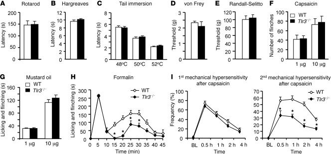
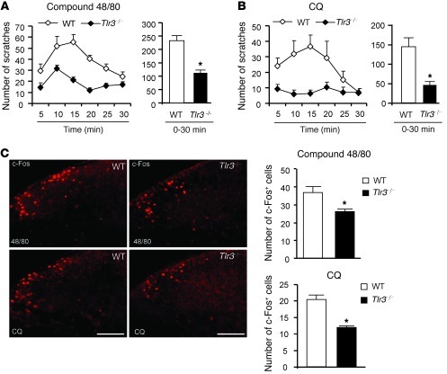
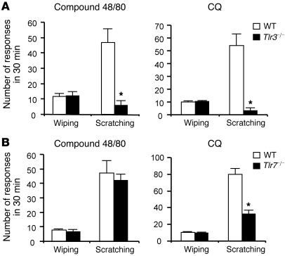
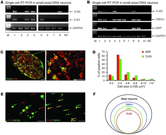
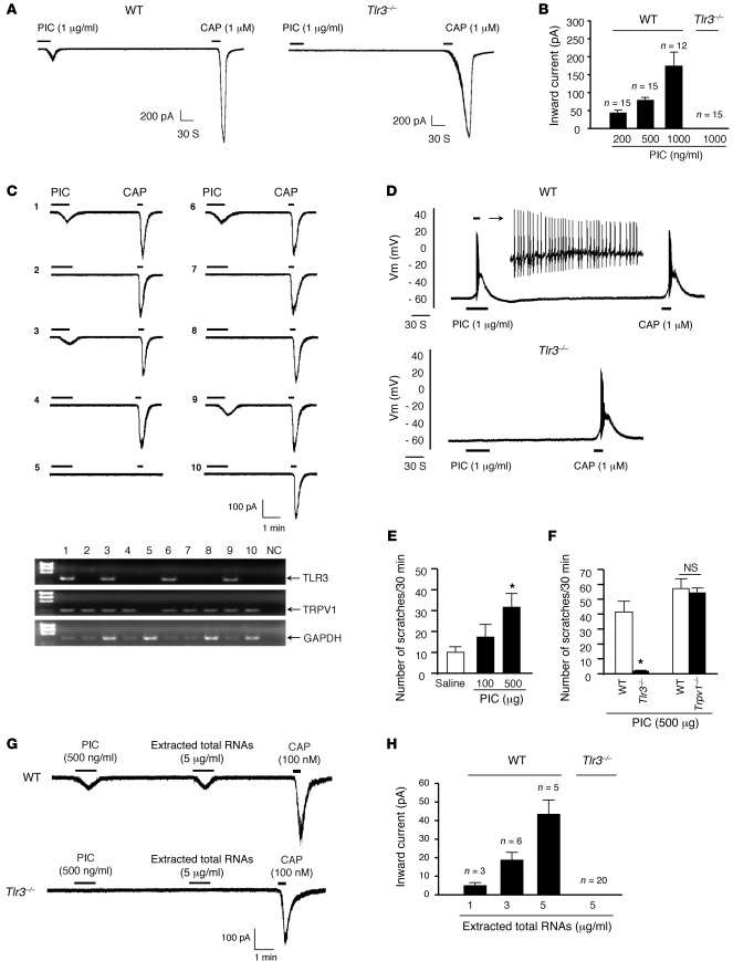
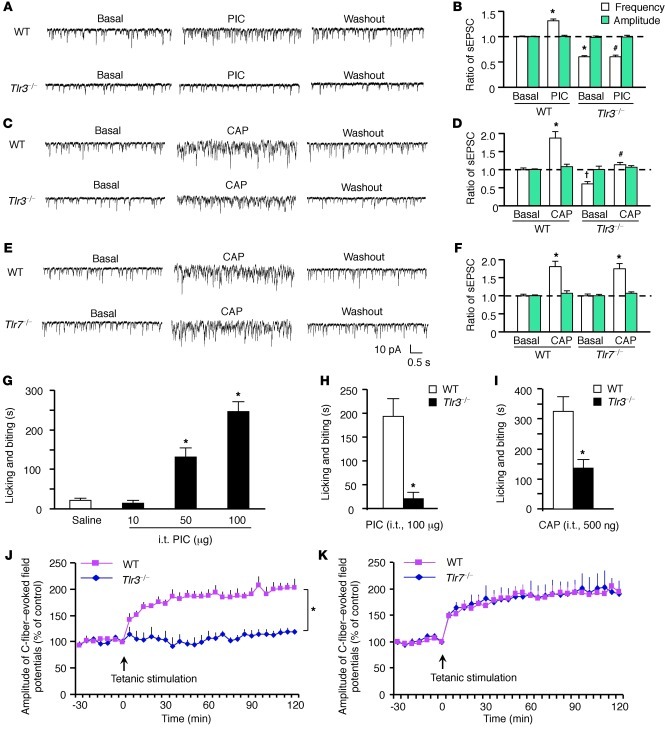
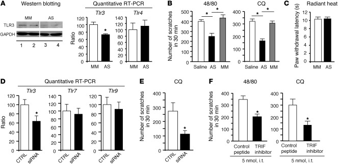
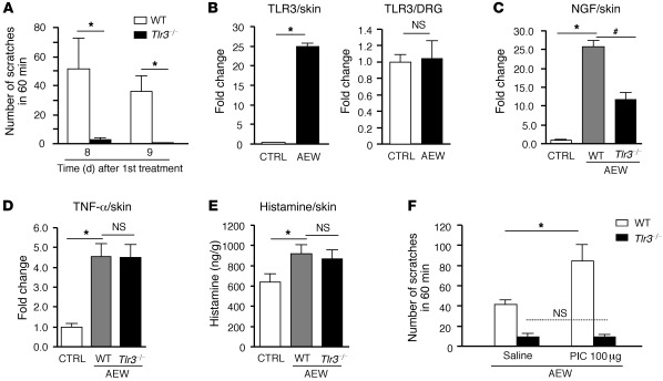
Itch, also known as pruritus, is a common, intractable symptom of several skin diseases, such as atopic dermatitis and xerosis. TLRs mediate innate immunity and regulate neuropathic pain, but their roles in pruritus are elusive. Here, we report that scratching behaviors induced by histamine-dependent and -independent pruritogens are markedly reduced in mice lacking the Tlr3 gene. TLR3 is expressed mainly by small-sized primary sensory neurons in dorsal root ganglions (DRGs) that coexpress the itch signaling pathway components transient receptor potential subtype V1 and gastrin-releasing peptide. Notably, we found that treatment with a TLR3 agonist induces inward currents and action potentials in DRG neurons and elicited scratching in WT mice but not Tlr3(-/-) mice. Furthermore, excitatory synaptic transmission in spinal cord slices and long-term potentiation in the intact spinal cord were impaired in Tlr3(-/-) mice but not Tlr7(-/-) mice. Consequently, central sensitization-driven pain hypersensitivity, but not acute pain, was impaired in Tlr3(-/-) mice. In addition, TLR3 knockdown in DRGs also attenuated pruritus in WT mice. Finally, chronic itch in a dry skin condition was substantially reduced in Tlr3(-/-) mice. Our findings demonstrate a critical role of TLR3 in regulating sensory neuronal excitability, spinal cord synaptic transmission, and central sensitization. TLR3 may serve as a new target for developing anti-itch treatment.


Figures

Figure 1
Figure 1. Intact acute pain but impaired central sensitization in Tlr3–/– mice.
(A) Motor function assessed by recording the falling latency in a Rotarod test of WT and Tlr3–/– mice. (B and C) Thermal sensitivity, measured by (B) Hargreaves and (C) tail immersion test, is comparable in WT and Tlr3–/– mice. (D and E) Mechanical sensitivity, assessed by (D) von Frey test and (E) Randall-Selitto test, is indistinguishable in WT and Tlr3–/– mice. (F and G) Acute spontaneous pain assessed by the number of flinches or duration of licking and flinching behaviors after intraplantar injection of (F) capsaicin and (G) mustard oil in WT and Tlr3–/– mice. (H) Spontaneous pain in the second phase (10–45 minutes) but not in the first phase (0–10 minutes) in the formalin test is decreased in Tlr3–/– mice. (I) Primary and secondary mechanical hypersensitivity after intraplantar injection of capsaicin (5 μg), assessed by percentage response (frequency) to a von Frey filament (0.16 g), in WT and Tlr3–/– mice. Tlr3–/– mice have intact primary mechanical hyperalgesia but impaired secondary mechanical hyperalgesia. BL, baseline. *P < 0.05, compared with WT mice, Student’s t test; n = 5–9 mice for each group. All the data are mean ± SEM.
Figure 2
Figure 2. Impaired scratching behaviors and reduced c-Fos expression in the spinal cords in Tlr3–/– mice.
(A and B) Scratches in every 5 minutes (left) and 0–30 minutes (right) induced by intradermal injection of 50 μl compound 48/80 (100 μg) and CQ (200 μg). Note a reduction of both histaminergic (compound 48/80) and nonhistaminergic (CQ) itch in Tlr3–/– mice. *P < 0.05, Student’s t test; n = 11–13 mice for each group. Mean ± SEM. Two-way repeated-measures ANOVA analysis also shows a significant difference in the time course of compound 48/80– and CQ-induced scratching between the 2 groups (P < 0.05). (C) c-Fos–like immunoreactivity in the dorsal horn of the cervical spinal cord in WT and Tlr3–/– mice 2 hours after intradermal injection of compound 48/80 (48/80) or CQ. Right panels show the number of c-Fos–positive neurons in the dorsal horn. Scale bars, 100 μm. *P < 0.05, Student’s t test; n = 4–6 mice. All the data are mean ± SEM.
Figure 3
Figure 3. Distinct role of TLRs in regulating pain and itch in a mouse cheek model.
Intradermal injection of compound 48/80 (50 μg) and CQ (100 μg) in the cheek induces either pain-like wiping by forelimbs or itch-like scratching by the hind limb. (A) Substantial reduction of compound 48/80– and CQ-induced scratching behaviors in Tlr3–/– mice. (B) CQ-induced but not compound 48/80–induced scratching is reduced in Tlr7–/– mice. Note that wiping behavior induced by compound 48/80 or CQ is normal in both Tlr3–/– and Tlr7–/– mice. *P < 0.05, Student’s t test; n = 5–8 mice. All the data are mean ± SEM.
Figure 4
Figure 4. Expression of TLR3 in a subset of small-sized DRG neurons.
(A) Single-cell RT-PCR analysis from dissociated small-sized DRG neurons showing the distinct and overlapped distribution patterns of TLR3 and TLR7 in DRG neurons. The lanes were run on the same gel but were noncontiguous. M, marker; NC, negative control. (B) Single-cell RT-PCR analysis from dissociated small-sized DRG neurons showing colocalization of TLR3 with TPRV1 and GRP. Similar results were obtained from 3 independent experiments in 30 cells collected from different animals. (C) Double immunostaining in DRGs showing co-colocalization of TLR3 and GRP. Red and yellow arrows indicate GRP+ only and double-labeled neurons, respectively. Scale bars: 50 μm. (D) Cell size distribution frequency of TLR3+ and GRP+ neurons. (E) Double immunostaining in cultured DRG neurons showing co-colocalization of TLR3 with TRPV1 but not with NF200. Green arrows indicate NF200+ or TRPV1+ neurons, red arrows indicate TLR3+ neurons, and yellow allows indicate double-labeled neurons. Scale bars: 50 μm. (F) A Venn diagram showing the relationship of TLR3+, GRP+, and TRPV1+ populations in a DRG. Note that all TLR3+ cells also express GRP and TRPV1.
Figure 5
Figure 5. PIC induces inward current and action potentials in dissociated DRG neurons and elicits scratching in WT mice via TLR3 activation.
(A) Inward currents evoked by PIC and capsaicin (CAP) in dissociated small-sized DRG neurons from WT and Tlr3–/– mice. Note that PIC fails to induce inward currents in Tlr3–/– mice. (B) Dose-dependent inward currents induced by PIC. The number of responsive neurons is indicated on the top of each bar. (C) A combination of patch-clamp recording and single-cell RT-PCR in small-sized DRG neurons shows that all 4 neurons (out of 10) that respond to PIC (200 ng/ml) also express Tlr3 mRNA. (D) Action potentials evoked by PIC and capsaicin in DRG neurons from WT and Tlr3–/– mice. Note that PIC does not induce action potentials in Tlr3–/– mice (n = 18 neurons). (E) Intradermal PIC induces dose-dependent scratching in WT mice. *P < 0.05, compared with vehicle; n = 6 mice. (F) Intradermal PIC induces scratching in WT mice but not Tlr3–/– mice. *P < 0.05, compared with WT, Student’s t test; n = 6 mice. (G) Inward currents evoked by the extracted total RNAs, PIC, and capsaicin in small-sized DRG neurons from WT and Tlr3–/– mice. (H) Dose-dependent inward currents induced by the total RNAs. The number of responsive neurons is indicated on the top of each bar. All the data are mean ± SEM.
Figure 6
Figure 6. Impaired synaptic transmission and LTP induction in the spinal cord dorsal horn of Tlr3–/– mice but not Tlr7–/– mice.
(AF) Patch-clamp recording of sEPSCs in lamina II neurons in spinal cord slices. (A, C, and E) Traces of sEPSCs in spinal cord slices of WT and Tlr3–/– mice after (A) PIC (100 ng/ml) and (C) capsaicin (1 μM) treatment and (E) of WT and Tlr7–/– mice after capsaicin treatment (1 μM). (B, D, and F) sEPSC frequency and amplitude after the same treatments in A, C, and E. PIC and capsaicin only increase the frequency but not amplitude of sEPSC frequency in WT mice but not in Tlr3–/– mice. *P < 0.05, P < 0.05, compared with WT pretreatment baseline; #P < 0.05, compared with WT-PIC or WT-capsaicin group, Student’s t test; n = 5–6 neurons. (G) Intrathecal (i.t.) PIC induces dose-dependent licking and biting in WT mice. *P < 0.05, Student’s t test, compared with vehicle; n = 6 mice. (H) Intrathecal PIC or (I) capsaicin induces licking and biting in WT mice but not Tlr3–/– mice. *P < 0.05, Student’s t test, compared with WT, n = 6 mice. (J and K) In vivo recordings of LTP of C-fiber–evoked filed potentials in the spinal cords of (J) Tlr3–/– and (K) Tlr7–/– mice and their corresponding WT controls. Note that spinal LTP is induced in Tlr7–/– mice but not in Tlr3–/– mice. *P < 0.05, compared with WT mice, 2-way repeated-measures ANOVA; n = 5 mice. All the data are mean ± SEM.
Figure 7
Figure 7. TLR3 signaling in DRGs of WT adult mice is required for pruritus.
(A) Knockdown of TLR3 expression in DRGs after intrathecal injections of Tlr3 AS-ODNs (10 μg daily for 5 days). The lanes ran on the same gel but were noncontiguous. Note that both TLR3 protein and mRNA levels but not Tlr4 mRNA levels in DRGs are decreased after AS-ODN treatment, revealed by Western blotting or quantitative PCR. *P < 0.05, Student’s t test; n = 4 mice. (B) Inhibition of compound 48/80– and CQ-induced scratching after treatment with TLR3 AS-ODNs. MM, mismatch oligodeoxynucleotides. *P < 0.05, Student t test; n = 5 mice. (C) No effects of TLR3 AS-ODNs on basal heat sensitivity (n = 5 mice). (D) Quantitative PCR reveals knockdown of Tlr3 but not Tlr7 and Tlr9 mRNA expression in DRGs after intrathecal injections of Tlr3-targeting siRNA (3 μg daily for 3 days). *P < 0.05, Student’s t test; n = 5 mice. (E) Inhibition of CQ-induced scratching after siRNA treatment. *P < 0.05, Student t test; n = 5 mice. (F) Intrathecal inhibition of TLR3 signaling with a peptide inhibitor of TRIF reduces scratching in mice. *P < 0.05, Student’s t test; n = 5 mice. All the data are mean ± SEM.
Figure 8
Figure 8. TLR3 is essential for dry skin–induced chronic itch.
(A) Spontaneous scratching induced by acetone and diethyether (1:1) following by water (AEW, twice a day for 7 days) on day 8 and 9 in WT and Tlr3–/– mice. *P < 0.05. (B) Real-time quantitative RT-PCR analysis showing TLR3 upregulation in skin but not DRGs of WT mice after AEW treatment. *P < 0.05. (C and D) Real-time quantitative RT-PCR analysis showing that AEW-induced upregulation of (C) NGF but not (D) TNF-α in the dry skin is abrogated in Tlr3–/– mice. *P < 0.05, #P < 0.05. (E) ELISA analysis showing that AEW-induced upregulation of histamine in skin is not altered in Tlr3–/– mice. *P < 0.05, compared with vehicle control (CTRL). (F) Intradermal injection of PIC enhances AEW-induced spontaneous scratching, which is abrogated in Tlr3–/– mice. *P < 0.05, Student’s t test; n = 5 mice in all cases. All the data are mean ± SEM.

Similar articles

Cited by

References

    1. Ikoma A, Steinhoff M, Stander S, Yosipovitch G, Schmelz M. The neurobiology of itch. Nat Rev Neurosci. 2006;7(7):535–547. doi: 10.1038/nrn1950. - DOI - PubMed
    1. Yosipovitch G, Greaves MW, Schmelz M. Itch. Lancet. 2003;361(9358):690–694. doi: 10.1016/S0140-6736(03)12570-6. - DOI - PubMed
    1. Paus R, Schmelz M, Biro T, Steinhoff M. Frontiers in pruritus research: scratching the brain for more effective itch therapy. J Clin Invest. 2006;116(5):1174–1186. doi: 10.1172/JCI28553. - DOI - PMC - PubMed
    1. Kawai T, Akira S. The role of pattern-recognition receptors in innate immunity: update on Toll-like receptors. Nat Immunol. 2010;11(5):373–384. doi: 10.1038/ni.1863. - DOI - PubMed
    1. Town T, Jeng D, Alexopoulou L, Tan J, Flavell RA. Microglia recognize double-stranded RNA via TLR3. J Immunol. 2006;176(6):3804–3812. - PubMed

Publication types

MeSH terms