. 2012 Jun;13(6):561-8.
doi: 10.1038/embor.2012.58.

CDK targeting of NBS1 promotes DNA-end resection, replication restart and homologous recombination

Affiliations

CDK targeting of NBS1 promotes DNA-end resection, replication restart and homologous recombination

Jacob Falck et al. EMBO Rep. 2012 Jun.

Abstract

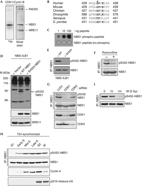
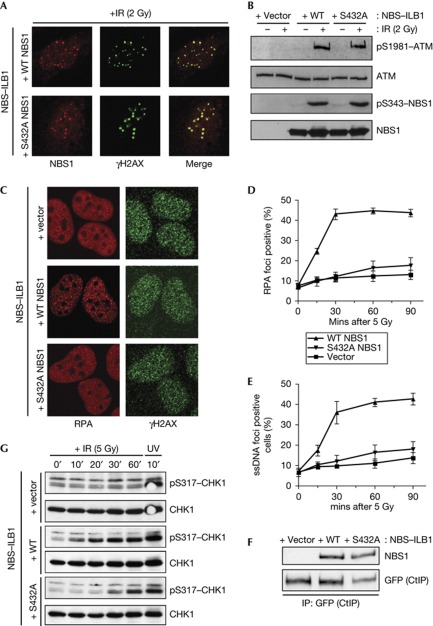
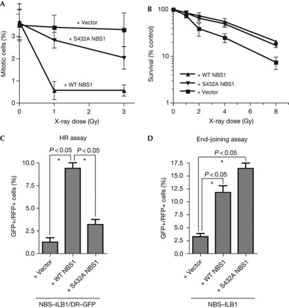
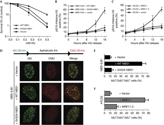
The conserved MRE11–RAD50–NBS1 (MRN) complex is an important sensor of DNA double-strand breaks (DSBs) and facilitates DNA repair by homologous recombination (HR) and end joining. Here, we identify NBS1 as a target of cyclin-dependent kinase (CDK) phosphorylation. We show that NBS1 serine 432 phosphorylation occurs in the S, G2 and M phases of the cell cycle and requires CDK activity. This modification stimulates MRN-dependent conversion of DSBs into structures that are substrates for repair by HR. Impairment of NBS1 phosphorylation not only negatively affects DSB repair by HR, but also prevents resumption of DNA replication after replication-fork stalling. Thus, CDK-mediated NBS1 phosphorylation defines a molecular switch that controls the choice of repair mode for DSBs.


Conflict of interest statement

The authors declare that they have no conflict of interest.

Figures

Figure 1
Figure 1
NBS1 Ser 432 is a CDK substrate. (A) Biochemical kinase assay with recombinant CDK1/CyclinB and purified MRN as substrate. (B) Alignment of region surrounding putative phospho-site in NBS1 orthologues. (C) Characterization of the NBS1–Ser 432 phospho-specific antibody. (D) NBS1 phospho-Ser 432 antibody recognizes NBS1 in whole-cell extracts and (E) after immunoprecipitation. (F) U2OS cells were treated or mock-treated with 50 μM roscovitine for 4 h. (G) Depletion of CDK1 or CDK2 in U2OS cells reduces NBS1 Ser 432 phosphorylation. (H) NBS1 Ser 432 phosphorylation is cell-cycle regulated. (I) NBS1 Ser 432 phosphorylation is reduced after DNA-damage induction. CDK, cyclin-dependent kinase; IP, immunoprecipitation; IR, ionizing radiation; MRN, MRE11–RAD50–NBS1; NBS, Nijmegen breakage syndrome; siRNA, small-interfering RNA; WT, wild-type.
Figure 2
Figure 2
NBS1 S432A impairs DNA-end resection. (A) IR-induced focus formation in complemented NBS cells. Cells were collected 1 h after irradiation. (B) Immunoblots from complemented NBS cells after irradiation. Cells were collected 1 h after irradiation. (C) IR-induced RPA foci from complemented NBS cells. Cells were irradiated with 5 Gy IR and collected 1 h after irradiation. (D,E): Quantification of IF data. At least 100 cells were counted per condition. Error bars represent standard deviations. (F) NBS1 coimmunoprecipitates with CtIP regardless of S432A mutation. NBS cells were transfected with a plasmid containing GFP–CtIP. (G) Kinetics of IR-induced CHK1 phosphorylation in complemented NBS cells. Cells were irradiated with 5 J/m2 ultraviolet. GFP, green fluorescent protein; IF, immunofluorescence; IP, immunoprecipitation; IR, ionizing radiation; NBS, Nijmegen breakage syndrome; RPA, replication protein A; ssDNA, single-stranded DNA; UV, ultraviolet light; WT, wild-type.
Figure 3
Figure 3
NBS1 S432A impairs HR without significantly affecting end joining. (A) G2/M assay in complemented NBS cells. Cells were collected 1 h after irradiation and measured for H3 pSer10 staining by flow cytometry. (B) IR survival. (C) HR assay in NBS/DR–GFP cells. (D) End-joining assay in complemented NBS cells. All quantitative data represent the average of three independent experiments (±s.d.). (*) Denotes statistically significant differences (P<0.05, unpaired t-test). HR, homologous recombination; GFP, green fluorescent protein; IR, ionizing radiation; NBS, Nijmegen breakage syndrome; RFP, red fluorescent protein; WT, wild-type.
Figure 4
Figure 4
NBS1 S432A impairs replication restart. (A) Cells were treated for 24 h with the indicated HU dose. Data represent the average of three independent experiments (±s.d.). (B) Mitotic entry after HU release in complemented NBS and (C) ATLD2 cells. Cells were treated with 2 mM HU for 24 h. Data represent the average of three independent experiments (±s.d.). (D) Replication restart after aphidicolin treatment in complemented NBS cells. (E) Quantification of IF data in complemented NBS and (F) ATLD2 cells. At least 100 cells were counted per condition. Error bars represent standard deviations. ATLD2, Ataxia telangiectasia-like disease; CldU, 5-chloro-2-deoxyuridine; HR, homologous recombination; HU, hydroxyurea; IdU, 5-iodo-2-deoxyuridine; IF, immunofluorescence; IR, ionizing radiation; NBS, Nijmegen breakage syndrome; RPA, replication protein A; UV, ultraviolet light; WT, wild-type.

References

    1. Jackson SP, Bartek J (2009) The DNA-damage response in human biology and disease. Nature 461: 1071–1078 - PMC - PubMed
    1. Harper JW, Elledge SJ (2007) The DNA damage response: ten years after. Mol Cell 28: 739–745 - PubMed
    1. Uziel T et al. (2003) Requirement of the MRN complex for ATM activation by DNA damage. EMBO J 22: 5612–5621 - PMC - PubMed
    1. Falck J, Coates J, Jackson SP (2005) Conserved modes of recruitment of ATM, ATR and DNA-PKcs to sites of DNA damage. Nature 434: 605–611 - PubMed
    1. Matsuoka S et al. (2007) ATM and ATR substrate analysis reveals extensive protein networks responsive to DNA damage. Science 316: 1160–1166 - PubMed

Publication types

MeSH terms