. 2012 Jun 5;125(22):2698-706.
doi: 10.1161/CIRCULATIONAHA.112.094714. Epub 2012 May 7.

Ranolazine decreases mechanosensitivity of the voltage-gated sodium ion channel Na(v)1.5: a novel mechanism of drug action

Affiliations

Ranolazine decreases mechanosensitivity of the voltage-gated sodium ion channel Na(v)1.5: a novel mechanism of drug action

Arthur Beyder et al. Circulation. .

Abstract

Background: Na(V)1.5 is a mechanosensitive voltage-gated sodium-selective ion channel responsible for the depolarizing current and maintenance of the action potential plateau in the heart. Ranolazine is a Na(V)1.5 antagonist with antianginal and antiarrhythmic properties.

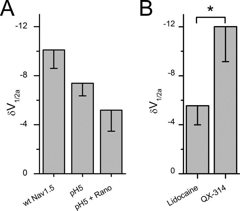
Methods and results: Mechanosensitivity of Na(V)1.5 was tested in voltage-clamped whole cells and cell-attached patches by bath flow and patch pressure, respectively. In whole cells, bath flow increased peak inward current in both murine ventricular cardiac myocytes (24±8%) and human embryonic kidney 293 cells heterologously expressing Na(V)1.5 (18±3%). The flow-induced increases in peak current were blocked by ranolazine. In cell-attached patches from cardiac myocytes and Na(V)1.5-expressing human embryonic kidney 293 cells, negative pressure increased Na(V) peak currents by 27±18% and 18±4% and hyperpolarized voltage dependence of activation by -11 mV and -10 mV, respectively. In human embryonic kidney 293 cells, negative pressure also increased the window current (250%) and increased late open channel events (250%). Ranolazine decreased pressure-induced shift in the voltage dependence (IC(50) 54 μmol/L) and eliminated the pressure-induced increases in window current and late current event numbers. Block of Na(V)1.5 mechanosensitivity by ranolazine was not due to the known binding site on DIVS6 (F1760). The effect of ranolazine on mechanosensitivity of Na(V)1.5 was approximated by lidocaine. However, ionized ranolazine and charged lidocaine analog (QX-314) failed to block mechanosensitivity.

Conclusions: Ranolazine effectively inhibits mechanosensitivity of Na(V)1.5. The block of Na(V)1.5 mechanosensitivity by ranolazine does not utilize the established binding site and may require bilayer partitioning. Ranolazine block of Na(V)1.5 mechanosensitivity may be relevant in disorders of mechanoelectric dysfunction.


Figures

Figure 1
Figure 1
Ranolazine blocks mechanosensitive response and peak currents of Na+ channels in murine cardiac myocytes and HEK cells transfected with NaV1.5. Left, A, Representative Na+ currents recorded by whole cell voltage-clamp from murine cardiac myocytes and B, HEK cells transfected with NaV1.5, elicited by stepping to -30 mV from -120 mV before (black traces, Flow OFF) or during (grey traces, Flow ON) bath flow, produced by rinsing solution through the recording chamber at 10 mL/min in the absence (Control, 0 μM) or presence (Ranolazine, 50 μM) of drug. Right, Average peak current densities in response to flow of solution without (filled symbols) or with (empty symbols) ranolazine (n=5; *P<0.05 compared to Flow OFF, †P<0.05 compared to 0 μM ranolazine, and P<0.05 interaction between flow and ranolazine blockade by two-way repeated measures ANOVA with Bonferroni multiple comparisons posttest).
Figure 2
Figure 2
Ranolazine blocks pressure-induced increase in peak current and hyperpolarizing shift of the voltage-dependence of activation and inactivation. A, B, C are controls and D, E, F are ranolazine (50 μM). A & D, single patch average Na+ currents elicited by stepping to -100 mV (dash dot) -50 mV (dot), -30 mV (solid) from -140 mV at 0 mmHg (black) and at -30 mmHg pressure (grey). First peak is activation and second peak is inactivation (availability). B & E, same patch difference currents (I-30 – I0) shown for -50 mV (dot black) and -30 mV (solid black). C & F, peak current-voltage (IV) for this patch at 0 mmHg (black) and -30 mmHg (grey), with activation (boxes) and inactivation (circles). Solid lines are Boltzmann fits of 0 mmHg (black) and -30 mmHg (grey).
Figure 3
Figure 3
Ranolazine blocks mechanosensitivity of NaV1.5 in a concentration-dependent manner. Bar graph is shift in the voltage dependence of activation (ΔV1/2a) for 0 μM ranolazine and scatter plot is ΔV1/2a versus increasing ranolazine concentrations. Solid line is a dose-response fit with IC50 of 53.6 μM.
Figure 4
Figure 4
Ranolazine inhibits pressure-induced increase in window current. A are controls and B are ranolazine (50 μM). A & B, single channel Na+ window current at HP -40 mV typical (black) and overlaid first fifty 300 msec traces (grey) at 0 mmHg and -30 mmHg (bracket). Single channel activity idealized before (black bar) and during (grey bar) the pressure pulse. Bar graphs show for -30 mmHg compared to 0 mmHg percent change of open channel event number (#30/#0 × 100) for control (Ai) and 50 μM ranolazine (Bi), and open channel lifetime (τ300 × 100) for control (Aii) and 50 μM ranolazine (Bii) (n=5, *P<0.05; paired t-test for events between 0 mmHg and -30mmHg).
Figure 5
Figure 5
Ranolazine inhibits pressure-induced increase in late open channel event number. A are controls and B are ranolazine (50 μM). A & B, single channel Na+ late current typical (black) and overlaid first fifty (grey) 200 msec long depolarizing pulses to 0 mV from HP -100 mV for ramp to -30 mmHg (bottom). Single channel activity in the last 100 msec of late current activity analyzed (grey bars). Bar graphs show for -30 mmHg compared to 0 mmHg percent change in open channel event number (#30/#0 × 100) for control (Ai) and 50 μM ranolazine (Bi), and for open channel lifetime (τ300 × 100) for control (Aii) and 50 μM ranolazine (Bii) (n=4, *P<0.05; paired t-test for events between 0 mmHg and -30mmHg).
Figure 6
Figure 6
Ranolazine blocks a mechanosensitive response but does not reduce peak currents of NaV1.5 F1760A expressed in HEK cells. Left, Na+ currents elicited by stepping to -30 mV from -120 mV before (black, Flow OFF) or during (grey, Flow ON) bath flow, produced by rinsing solution through the recording chamber at 10 mL/min in the absence (Control, 0 μM) or presence (Ranolazine, 50 μM) of drug. Right, Average peak current densities of NaV1.5 F1760A in response to flow of solution without (filled symbols) or with (empty symbols) 50 μM ranolazine (n=6; *P<0.05 compared to Flow OFF, P>0.05 compared to 0 μM ranolazine, and P>0.05 interaction between flow and ranolazine blockade by two-way repeated measures ANOVA with Bonferroni multiple comparisons posttest).
Figure 7
Figure 7
Ranolazine block of mechanosensitivity is independent of F1760. A, the fractional decrease in peak current [(Ictr-Irano)/Ictr] obtained from peak of IV curves in response to 50 μM ranolazine for wild-type NaV1.5 (left) and F1760A (right) (n=4; *P<0.05; two-sample t-test). B, mechanosensitivity of F1760A shown as a shift in half-point of voltage-dependence of activation (ΔV1/2a) without ranolazine and significantly reduced with 50 μM ranolazine (n=4-7, *P<0.05; two sample t-test).
Figure 8
Figure 8
Neutral ranolazine and lidocaine block mechanosensitivity of NaV1.5. A, In cell-attached patches, pressure-induced ΔV1/2a with wt NaV1.5 in Ringer solution at pH 7.4 was not statistically different from ΔV1/2a when the pipette solution contained Ringer solution at pH 5 (n=5-7, P>0.05; two-sample t-test). ΔV1/2a was also not statistically different for pH 5 solution and ranolazine (50 μM) in pH 5 solution (n=7, P>0.05; two-sample t-test). B, Shift in V1/2a with pressure with lidocaine (50 μM) is significantly less than with QX-314 (500 μM) (n=3-5, *P<0.05; two-sample t-test).

Comment in

References

    1. Cooper PJ, Lei M, Cheng LX, Kohl P. Selected contribution: Axial stretch increases spontaneous pacemaker activity in rabbit isolated sinoatrial node cells. J Appl Physiol. 2000;89:2099–2104. - PubMed
    1. Lerman BB, Burkhoff D, Yue DT, Franz MR, Sagawa K. Mechanoelectrical feedback: Independent role of preload and contractility in modulation of canine ventricular excitability. J Clin Invest. 1985;76:1843–1850. - PMC - PubMed
    1. Sung D, Mills RW, Schettler J, Narayan SM, Omens JH, McCulloch AD. Ventricular filling slows epicardial conduction and increases action potential duration in an optical mapping study of the isolated rabbit heart. J Cardiovasc Electrophysiol. 2003;14:739–749. - PubMed
    1. Maron BJ, Poliac LC, Kaplan JA, Mueller FO. Blunt impact to the chest leading to sudden death from cardiac arrest during sports activities. N Engl J Med. 1995;333:337–342. - PubMed
    1. Wang Z, Taylor LK, Denney WD, Hansen DE. Initiation of ventricular extrasystoles by myocardial stretch in chronically dilated and failing canine left ventricle. Circulation. 1994;90:2022–2031. - PubMed

Publication types

MeSH terms