. 2012 May 22;109(21):7974-81.
doi: 10.1073/pnas.1120210109. Epub 2012 May 7.

A common X-linked inborn error of carnitine biosynthesis may be a risk factor for nondysmorphic autism

Affiliations

A common X-linked inborn error of carnitine biosynthesis may be a risk factor for nondysmorphic autism

Patrícia B S Celestino-Soper et al. Proc Natl Acad Sci U S A. .

Abstract

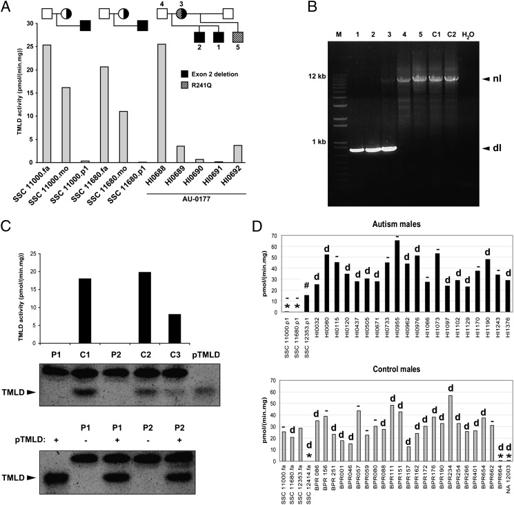
We recently reported a deletion of exon 2 of the trimethyllysine hydroxylase epsilon (TMLHE) gene in a proband with autism. TMLHE maps to the X chromosome and encodes the first enzyme in carnitine biosynthesis, 6-N-trimethyllysine dioxygenase. Deletion of exon 2 of TMLHE causes enzyme deficiency, resulting in increased substrate concentration (6-N-trimethyllysine) and decreased product levels (3-hydroxy-6-N-trimethyllysine and γ-butyrobetaine) in plasma and urine. TMLHE deficiency is common in control males (24 in 8,787 or 1 in 366) and was not significantly increased in frequency in probands from simplex autism families (9 in 2,904 or 1 in 323). However, it was 2.82-fold more frequent in probands from male-male multiplex autism families compared with controls (7 in 909 or 1 in 130; P = 0.023). Additionally, six of seven autistic male siblings of probands in male-male multiplex families had the deletion, suggesting that TMLHE deficiency is a risk factor for autism (metaanalysis Z-score = 2.90 and P = 0.0037), although with low penetrance (2-4%). These data suggest that dysregulation of carnitine metabolism may be important in nondysmorphic autism; that abnormalities of carnitine intake, loss, transport, or synthesis may be important in a larger fraction of nondysmorphic autism cases; and that the carnitine pathway may provide a novel target for therapy or prevention of autism.


Conflict of interest statement

Conflict of interest statement: Multiple authors are based in the Department of Molecular and Human Genetics at Baylor College of Medicine, which offers extensive genetic laboratory testing, and Baylor College of Medicine derives revenue from this activity. The department offers biochemical and molecular diagnostic testing for trimethyllysine hydroxylase, epsilon gene deficiency. P.B.S.C.-S., S.V., F.M.V., and A.L.B. have filed a patent related to some of the work reported.

Figures

Fig. 1.
Fig. 1.
Carnitine biosynthesis and homeostasis in humans. Carnitine is synthesized in four enzymatic steps. After release of TML by lysosomal protein degradation, this compound is hydroxylated by TMLD, producing HTML. HTML is cleaved by HTML aldolase (HTMLA) into TMABA and glycine. Subsequently, TMABA is oxidized by TMABA-dehydroxygenase (TMABA-DH) to form 4-N-trimethylaminobutyrate, also named γ-butyrobetaine (γBB). Finally, γBB is hydroxylated by γBBD, yielding l-carnitine. Because TMLD is located in mitochondria, TML needs to be transported out of the lysosome and across the inner mitochondrial membrane into the mitochondrial matrix by means of transporters, which are unknown at this time. Depending on the subcellular localization of the HTLMA (also uncertain, likely the cytosol), HTML or γBB needs to be transported back to the cytosol (where γBBD is located). In cells that do not contain γBBD, γBB is exported from the cell and imported into tissues (liver, kidney, and brain in humans) that do express γBBD by means of at least one specific transporter, presumably SLC6A13. Carnitine is transported by OCTN2 and other lower affinity transporters (not shown).
Fig. 2.
Fig. 2.
Exon 2-containing deletions found in TMLHE and structure of TMLHE. (A) Array CGH showing eight exonic deletions in TMLHE, with the relative location of TMLHE exons 2–6 aligned above the array CGH plots (GRCh37/hg19 assembly; http://genome.ucsc.edu). The horizontal axis shows chromosome position, and the vertical axis shows the log2 ratio of array signal. Semitransparent filled boxes on CGH plots highlight the regions of aberration; all samples have deletions involving exon 2, and most have a separate deletion in intron 1 (red circle). All samples are autism probands with identifiers found in Tables S1, S4, and S5. (B) Twenty-four unrelated individuals with deletions involving exon 2 of TMLHE are mapped. Deletion coordinates were determined by array CGH unless specified by PCR assay. White arrowheads indicate the continuation of the deletion for SSC 13928.p1. NA 12003 is an unaffected individual whose deletion is published (38) and was better characterized in this study. BPR 664 is an unaffected individual. (C) Diagram of gene structure. Large open arrows represent near-identical inverted repeats. Fa, father; P1, proband; HI, AGRE individuals; #, individual first described by Celestino-Soper et al. (10).
Fig. 3.
Fig. 3.
Genetic and enzymatic characterization of hemizygous deletion of exon 2. (A) TMLD activity measured in lymphoblast homogenates of three families with exon 2 deletion. (B) PCR assay results for the AU 0177 family showing the deletion in the two affected brothers (1, 2) and in the mother (3) but not in the father (4), unaffected maternal half-brother (5), or unaffected controls (C1 and C2). There is bias of amplification in the mother, such that the normal band is faint. dl, deletion; nl, normal. (C) (Upper) TMLD activity and Western blot analysis of 2 individuals with exon 2 deletion (P1, HI0690; P2, BPR664) and three controls (C1–C3). Purified TMLD (pTMLD) is used as a positive control. (Lower) Western blot analysis of 2 individuals (P1 and P2) with (+) or without (−) addition of pTMLD, showing the complete absence of protein in cases of exon 2 deletion and confirmation of the identity of the immunoreactive material as TMLD. The upper band in the Western blot is an irrelevant protein. (D) (Upper) TMLD activity measured in lymphoblast homogenates from several autism males. *,TMLHE exon 2 deletion; #, E287K; d, deletion in intron 1 in 13 individuals; −, no deletion in intron 1 in 9 individuals. SSC 12353.p1 was not tested for the presence of intron 1 deletion. (Lower) TMLD activity measured in lymphoblast homogenates from male controls. BPR indicates local unaffected controls, and NA 12003 is an unaffected individual. SSC 12353.fa was not tested for presence of intron 1 deletion. There was no apparent correlation of the level of enzyme activity with the presence or absence of the intronic deletion. For A, C, and D, assays were run in duplicate and the average is plotted without error bars. fa, father; mo, mother; p1, proband.
Fig. 4.
Fig. 4.
Increased TML and decreased HTML and γBB in patients with TMLHE exon 2 deletions. (A) Bar diagram of concentrations (mean ± SD) of carnitine biosynthesis intermediates in urine of two patients with exon 2 deletion (1, HI0690; 2, HI0691) compared with controls. (B) Box and whisker plot of carnitine biosynthesis intermediates in plasma of seven patients with exon 2 deletion (HI0690, HI0691, SSC 13928.p1, SSC 13489.p1, SSC 11000.p1, SSC 11229.p1, and SSC 11680.p1) compared with controls. (C) Box and whisker plot showing the diagnostic potential of the (HTML + γBB)/HTML ratio as an indicator of TMLHE deficiency. All seven patients have a very low ratio. The black square represents the mean of the controls, and whiskers show the minimum and maximum values of the control group.

References

    1. Conference Proceedings (2004) Carnitine. The science behind a conditionally essential nutrient. Proceedings of a conference. March 25–26, 2004. Bethesda, MD. Ann N Y Acad Sci 1033:1–197. - PubMed
    1. Flanagan JL, Simmons PA, Vehige J, Willcox MD, Garrett Q. Role of carnitine in disease. Nutr Metab (Lond) 2010;7:30. - PMC - PubMed
    1. Vaz FM, Wanders RJ. Carnitine biosynthesis in mammals. Biochem J. 2002;361:417–429. - PMC - PubMed
    1. Slonim AE, et al. Dietary-dependent carnitine deficiency as a cause of nonketotic hypoglycemia in an infant. J Pediatr. 1981;99:551–555. - PubMed
    1. Schmidt-Sommerfeld E, Penn D. Carnitine and total parenteral nutrition of the neonate. Biol Neonate. 1990;58(Suppl 1):81–88. - PubMed

Publication types

MeSH terms

LinkOut - more resources