. 2012 Jan 6:2:92.
doi: 10.3389/fimmu.2011.00092. eCollection 2011.

The human c1q globular domain: structure and recognition of non-immune self ligands

Affiliations

The human c1q globular domain: structure and recognition of non-immune self ligands

Christine Gaboriaud et al. Front Immunol. .

Abstract

C1q, the ligand-binding unit of the C1 complex of complement, is a pattern recognition molecule with the unique ability to sense an amazing variety of targets, including a number of altered structures from self, such as apoptotic cells. The three-dimensional structure of its C-terminal globular domain, responsible for its recognition function, has been solved by X-ray crystallography, revealing a tightly packed heterotrimeric assembly with marked differences in the surface patterns of the subunits, and yielding insights into its versatile binding properties. In conjunction with other approaches, this same technique has been used recently to decipher the mechanisms that allow this domain to interact with various non-immune self ligands, including molecules known to provide eat-me signals on apoptotic cells, such as phosphatidylserine and DNA. These investigations provide evidence for a common binding area for these ligands located in subunit C of the C1q globular domain, and suggest that ligand recognition through this area down-regulates C1 activation, hence contributing to the control of the inflammatory reaction. The purpose of this article is to give an overview of these advances which represent a first step toward understanding the recognition mechanisms of C1q and their biological implications.

Keywords: C1q; X-ray crystallography; complement; innate immunity; ligand recognition.


Figures

Figure 1
Figure 1
Structure of the C1q globular domain. (A) Top view and (B) side view of the heterotrimeric assembly. Subunits A, B, and C are shown in blue, green, and red, respectively. β-strands are labeled according to the tumor necrosis factor nomenclature, and the Ca2+ ion is represented as a golden sphere. N and C indicate the N- and C-terminal ends of each gC1q subunit. (C) Side view of the three-dimensional C1q model derived from the structure of the globular domain. The N-linked oligosaccharide attached to each globular domain is represented in yellow. (D) Structure of the gC1q subunits. Subunits A, B, and C are superimposed. Only the free cysteine and disulfide bond of subunit C are displayed for clarity. (Modified from Gaboriaud et al., 2003, 2004).
Figure 2
Figure 2
Surface properties of the C1q globular domain. (A–C) Side views of the heterotrimer seen from subunits A, B, and C, respectively. (D) Top view of the heterotrimer. The side chains of Arg, Lys, His, Asp, and Glu residues are shown in deep blue, light blue, green, red, and magenta, respectively. Hydrophobic residues are shown in yellow, and aromatic ones are in orange. The lines in (D) indicate the approximate subunit boundaries. (From Gaboriaud et al., 2003).
Figure 3
Figure 3
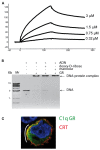
C1q binding to apoptotic cell ligands. (A) Surface plasmon resonance analysis of the binding of the C1q globular domain to immobilized phosphatidylserine. The concentrations of the C1q globular domain are indicated. (B) Electrophoretic mobility shift assay showing that complex formation between DNA and the C1q globular domain is partly inhibited by 100 mM deoxy-D-ribose. Mannose was used as a negative control. DNA molecular weight markers are shown on the left. (C) Early apoptotic HeLa cells were submitted to a double-immunofluorescence labeling for the C1q globular domain (C1q GR, green) and calreticulin (CRT, red) followed by confocal laser microscopy detection. Nuclei were labeled with Hoechst (blue). The white bow indicates areas where the C1q globular domain co-localizes with calreticulin. (From Païdassi et al., 2008a,b, 2011).
Figure 4
Figure 4
Location of three non-immune self ligand-binding sites in the C1q globular domain. (A) Overall view of the C1q globular domain indicating the relative positioning of the binding sites for heparan sulfate (HS), deoxy-D-ribose (dR), and phosphoserine (PS). (B–D) Detailed views of the binding sites for deoxy-D-ribose, heparan sulfate, and phosphoserine, respectively. Residues of the C subunit involved in the interaction are shown. In (C), the ligand portion displayed in sticks corresponds to the major interpretable extra electron-density. In (D), a double conformation was used to interpret the extra-density corresponding to phosphoserine. (Modified from Garlatti et al., and Païdassi et al., 2008a).
Figure 5
Figure 5
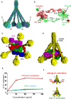
Implications on C1 activation of the location of ligand-binding sites in the C1q globular domain. (A) Location of the ligand-binding sites according to the current version of the C1q model. Nisl: non-immune self ligands binding area as described in Figure 4; IC: proposed binding sites for IgG-containing immune complexes. (B) Dimeric structure of the C1r catalytic domain in its “resting state.” In this configuration, a distance of 90 Å is observed between the catalytic site (C1r_cat) of one subunit and the activation site (colored star) to be cleaved in the other subunit. Such a distance prevents unwanted spontaneous C1r autoactivation, which requires transient disruption of the dimer. (C,D) Bottom and side views, respectively, of the current C1 model (Bally et al., ; Brier et al., 2010). In (C) white stars show the approximate positions of the C1r activation sites. (E) C1 activation by IgG-containing immune complexes, heparin and DNA. (F) Schematic interpretation of (E). According to our hypothesis, a strong outward movement of the C1q stems (red arrows) induced by binding to immune complexes is expected to disrupt the C1r catalytic dimer and thereby induce C1 activation. In contrast, binding to non-immune self ligands would generate little or no activation (green arrows). (Modified from Budayova-Spano et al., and Garlatti et al., 2010).

Similar articles

Cited by

References

    1. Bally I., Rossi V., Lunardi T., Thielens N. M., Gaboriaud C., Arlaud G. J. (2009). Identification of the C1q-binding sites of human C1r and C1s: a refined three-dimensional model of the C1 complex of complement. J. Biol. Chem. 284, 19340–1934810.1074/jbc.M109.004473 - DOI - PMC - PubMed
    1. Biro A., Ling W. L., Arlaud G. J. (2010). Complement protein C1q recognizes enzymatically modified low-density lipoprotein through unesterified fatty acids generated by cholesterol esterase. Biochemistry 49, 2167–217610.1021/bi9021022 - DOI - PubMed
    1. Biro A., Thielens N. M., Cervenak L., Prohaszka Z., Füst G., Arlaud G. J. (2007). Modified low density lipoproteins differentially bind and activate the C1 complex of complement. Mol. Immunol. 44, 1169–117710.1016/j.molimm.2006.06.013 - DOI - PubMed
    1. Bogin O., Kvansakul M., Rom E., Singer J., Yayon A., Hohenester E. (2002). Insights into Schmid metaphyseal chondrodysplasia from the crystal structure of the collagen X NC1 domain trimer. Structure 10, 165–17310.1016/S0969-2126(02)00697-4 - DOI - PubMed
    1. Botto M., Walport M. J. (2002). C1q, autoimmunity and apoptosis. Immunobiology 205, 395–40610.1078/0171-2985-00141 - DOI - PubMed

LinkOut - more resources