. 2012 Mar 5:3:40.
doi: 10.3389/fimmu.2012.00040. eCollection 2012.

Escape from the Phagosome: The Explanation for MHC-I Processing of Mycobacterial Antigens?

Affiliations

Escape from the Phagosome: The Explanation for MHC-I Processing of Mycobacterial Antigens?

Melanie J Harriff et al. Front Immunol. .

Abstract

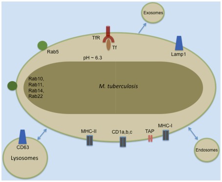
Mycobacterium tuberculosis (Mtb) is thought to live in an altered phagosomal environment. In this setting, the mechanisms by which mycobacterial antigens access the major histocompatibility class I (MHC-I) processing machinery remain incompletely understood. There is evidence that Mtb antigens can be processed in both endocytic and cytosolic environments, with different mechanisms being proposed for how Mtb antigens can access the cytosol. Recently, electron microscopy was used to demonstrate that Mtb has the potential to escape the phagosome and reside in the cytosol. This was postulated as the primary mechanism by which Mtb antigens enter the MHC-I processing and presentation pathway. In this commentary, we will review data on the escape of Mtb from the cytosol and whether this escape is required for antigen presentation to CD8(+) T cells.

Keywords: CD8+ T cells; MHC-I antigen processing; Mycobacterium tuberculosis; phagosome.


Figures

Figure 1
Figure 1
Common markers of the Mtb phagosome.
Figure 2
Figure 2
Cytosolic and vacuolar pathways for processing and presenting Mtb antigens in the context of MHC-I.

References

    1. Abdallah A. M., Gey Van Pittius N. C., Champion P. A., Cox J., Luirink J., Vandenbroucke-Grauls C. M., Appelmelk B. J., Bitter W. (2007). Type VII secretion – mycobacteria show the way. Nat. Rev. Microbiol. 5, 883–89110.1038/nrmicro1773 - DOI - PubMed
    1. Ackerman A. L., Kyritsis C., Tampe R., Cresswell P. (2003). Early phagosomes in dendritic cells form a cellular compartment sufficient for cross presentation of exogenous antigens. Proc. Natl. Acad. Sci. U.S.A. 100, 12889–1289410.1073/pnas.1735556100 - DOI - PMC - PubMed
    1. Amigorena S., Savina A. (2010). Intracellular mechanisms of antigen cross presentation in dendritic cells. Curr. Opin. Immunol. 22, 109–11710.1016/j.coi.2010.01.022 - DOI - PubMed
    1. Armstrong J. A., Hart P. D. (1971). Response of cultured macrophages to Mycobacterium tuberculosis, with observations on fusion of lysosomes with phagosomes. J. Exp. Med. 134, 713–74010.1084/jem.134.3.713 - DOI - PMC - PubMed
    1. Bach H., Papavinasasundaram K. G., Wong D., Hmama Z., Av-Gay Y. (2008). Mycobacterium tuberculosis virulence is mediated by PtpA dephosphorylation of human vacuolar protein sorting 33B. Cell Host Microbe 3, 316–32210.1016/j.chom.2008.03.008 - DOI - PubMed