

doi: 10.1155/2012/578721.
Epub 2012 Apr 11.
Wavelet transform fuzzy algorithms for dermoscopic image segmentation
Affiliations
- PMID: 22567042
- PMCID: PMC3332176
- DOI: 10.1155/2012/578721
Item in Clipboard
Wavelet transform fuzzy algorithms for dermoscopic image segmentation
Comput Math Methods Med.
2012.
Abstract
This paper presents a novel approach to segmentation of dermoscopic images based on wavelet transform where the approximation coefficients have been shown to be efficient in segmentation. The three novel frameworks proposed in this paper, W-FCM, W-CPSFCM, and WK-Means, have been employed in segmentation using ROC curve analysis to demonstrate sufficiently good results. The novel W-CPSFCM algorithm permits the detection of a number of clusters in automatic mode without the intervention of a specialist.
Figures
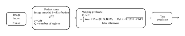
Block diagram of statistical region merging.
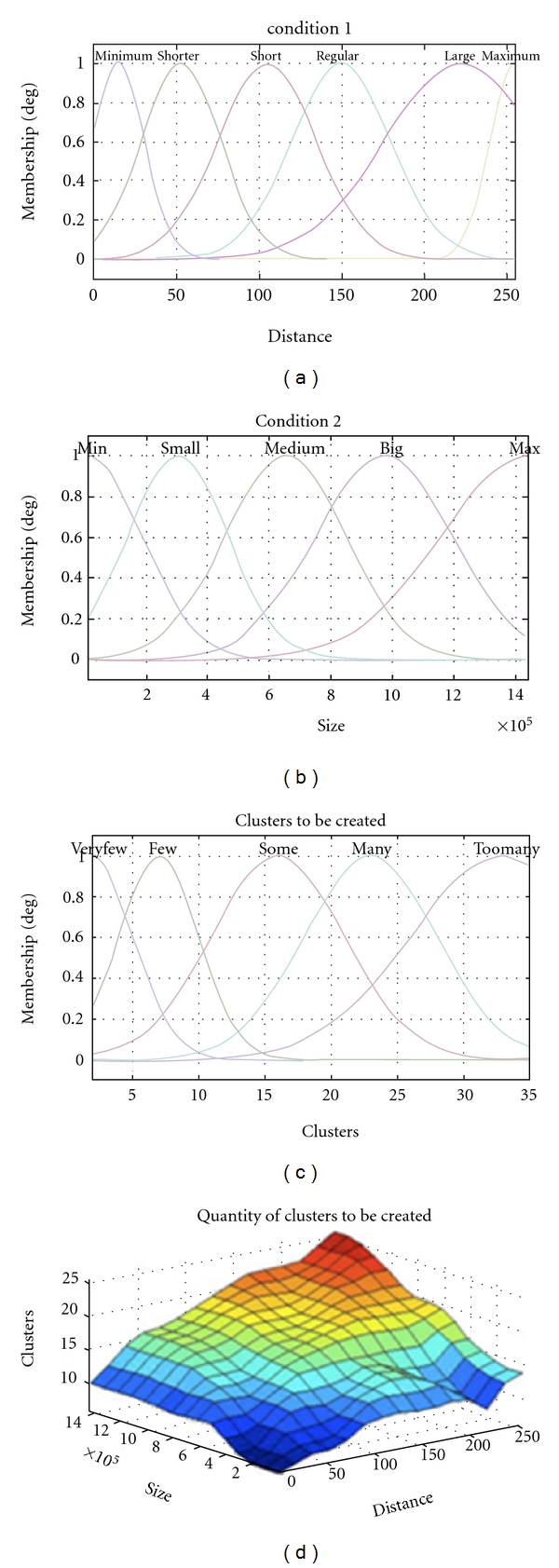
Preselection of the number of clusters.
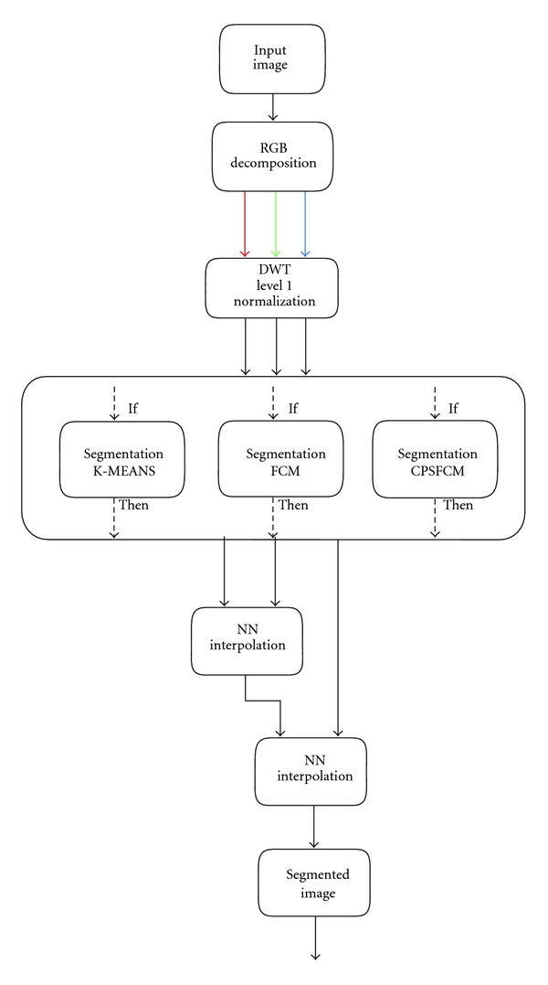
Block diagram of the proposed algorithms: WK-MEANS, W-FCM, and W-CPSFCM.
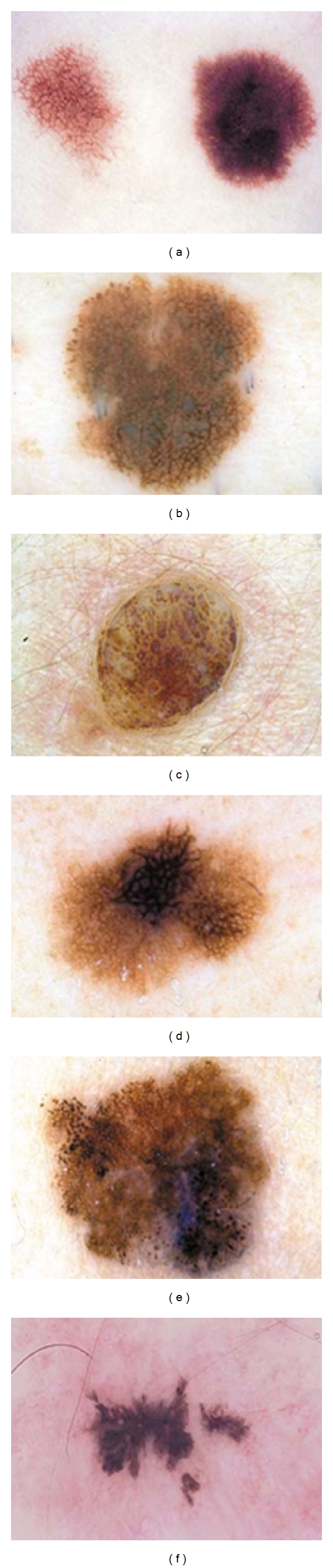
Dermoscopic images used in this study: (a) Clark's nevus (lesion 1), (b) Clark nevus's (lesion 2), (c) dermal nevus (lesion 3), (d) melanoma (lesion 4), (e) melanoma (lesion 5), (f) recurrent nevus (lesion 6).
GT of dermoscopic images used in this study: (a) Clark nevus's (lesion 1), (b) Clark's nevus (lesion 2), (c) dermal nevus (lesion 3), (d) melanoma (lesion 4), (e) melanoma (lesion 5), (f) recurrent nevus (lesion 6).

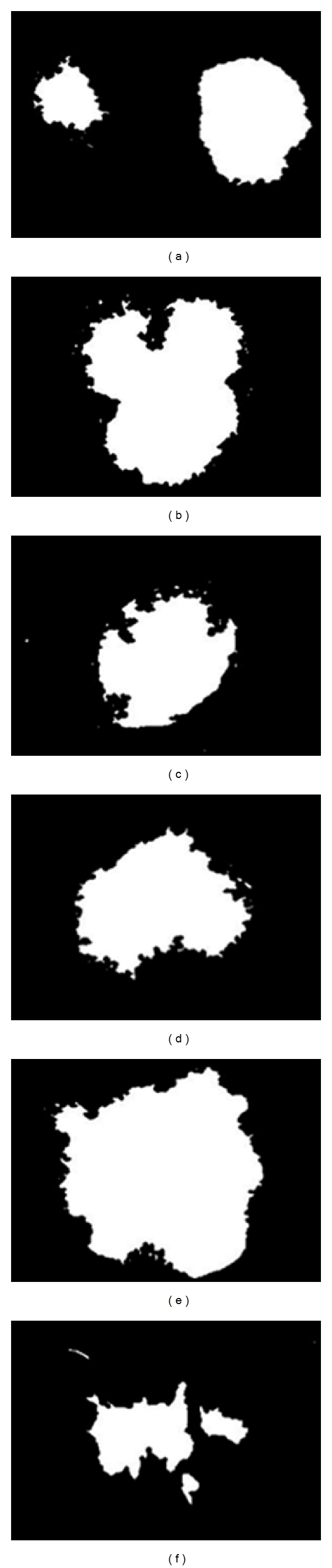
Image segmentation results under different algorithms using (a) melanoma, (b) ground truth, (c) FCM, (d) W-FCM with WF Coiflets 3, (e) W-FCM with Daubechies 4, (f) W-FCM with WF biorthogonal 6.8, (g) W-FCM with WAF up2, (h) W-FCM with WAF π
6, and (i) W-FCM with WAF fup2, (j) W-FCM with WAF e
2.
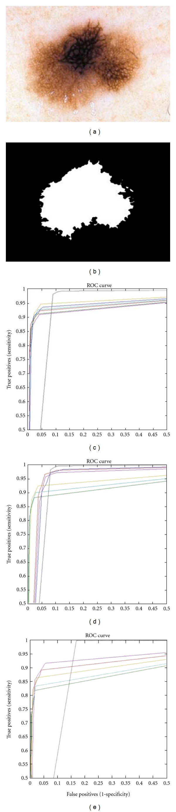
(a) Lesion 4 melanoma, (b) ground truth image; ROC curves for (c) WK-Means algorithm, (d) FCM algorithm, and (e) W-CPSFCM: for WF Daubechies 4 (dark blue), for WF biorthogonal 6.8 (red), for WF Coiflets 3 (purple), for WAF up2 (dark green), for WAF fup2 (aqua), and for WAF π
6 (light green); FCM (black).
References
-
- Argenziano G, Soyer HP. Dermoscopy of pigmented skin lesions—a valuable tool for early diagnosis of melanoma. Lancet Oncology. 2001;2(7):443–449. - PubMed
-
- Vestergaard ME, Macaskill P, Holt PE, Menzies SW. Dermoscopy compared with naked eye examination for the diagnosis of primary melanoma: a meta-analysis of studies performed in a clinical setting. British Journal of Dermatology. 2008;159(3):669–676. - PubMed
-
- Argenziano G, Fabbrocini G, Carli P, de Giorgi V, Sammarco E, Delfino M. Epiluminescence microscopy for the diagnosis of doubtful melanocytic skin lesions: comparison of the ABCD rule of dermatoscopy and a new 7-point checklist based on pattern analysis. Archives of Dermatology. 1998;134(12):1563–1570. - PubMed
-
- Ascierto PA, Palmieri G, Celentano E, et al. Sensitivity and specificity of epiluminescence microscopy: evaluation on a sample of 2731 excised cutaneous pigmented lesions. British Journal of Dermatology. 2000;142(5):893–898. - PubMed
Publication types
MeSH terms
LinkOut - more resources
Full Text Sources
Medical
