

Transcript profiling identifies dynamic gene expression patterns and an important role for Nrf2/Keap1 pathway in the developing mouse esophagus
- PMID: 22567161
- PMCID: PMC3342176
- DOI: 10.1371/journal.pone.0036504
Transcript profiling identifies dynamic gene expression patterns and an important role for Nrf2/Keap1 pathway in the developing mouse esophagus
Erratum in
- PLoS One. 2012;7(5): doi/10.1371/annotation/b2554ac2-5eec-4fc1-b625-c79f0cc064ed
Abstract
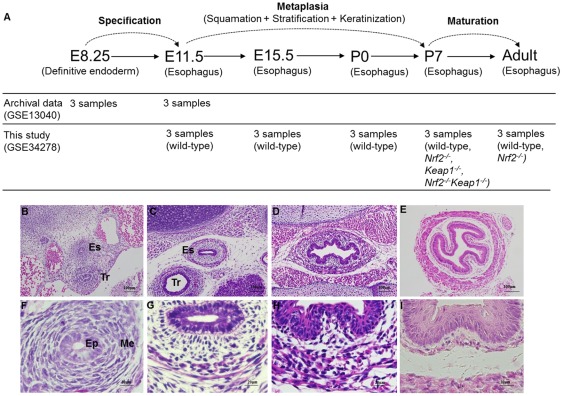
Background and aims: Morphological changes during human and mouse esophageal development have been well characterized. However, changes at the molecular level in the course of esophageal morphogenesis remain unclear. This study aims to globally profile critical genes and signaling pathways during the development of mouse esophagus. By using microarray analysis this study also aims to determine how the Nrf2/Keap1 pathway regulates the morphogenesis of the esophageal epithelium.
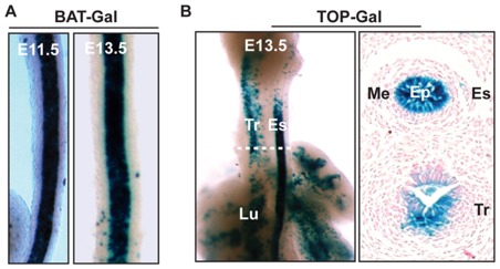
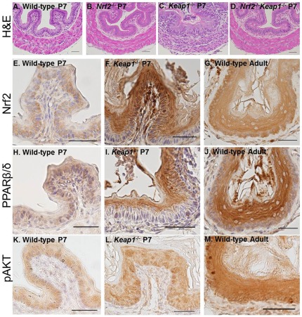
Methods: Gene expression microarrays were used to survey gene expression in the esophagus at three critical phases: specification, metaplasia and maturation. The esophagi were isolated from wild-type, Nrf2(-/-), Keap1(-/-), or Nrf2(-/-)Keap1(-/-) embryos or young adult mice. Array data were statistically analyzed for differentially expressed genes and pathways. Histochemical and immunohistochemical staining were used to verify potential involvement of the Wnt pathway, Pparβ/δ and the PI3K/Akt pathway in the development of esophageal epithelium.
Results: Dynamic gene expression patterns accompanied the morphological changes of the developing esophagus at critical phases. Particularly, the Nrf2/Keap1 pathway had a baseline activity in the metaplasia phase and was further activated in the maturation phase. The Wnt pathway was active early and became inactive later in the metaplasia phase. In addition, Keap1(-/-) mice showed increased expression of Nrf2 downstream targets and genes involved in keratinization. Microarray and immunostaining data also suggested that esophageal hyperkeratosis in the Keap1(-/-) mice was due to activation of Pparβ/δ and the PI3K/Akt pathway.
Conclusions: Morphological changes of the esophageal epithelium are associated with dynamic changes in gene expression. Nrf2/Keap1 pathway activity is required for maturation of mouse esophageal epithelium.
Conflict of interest statement
Figures
References
MeSH terms
Substances
Associated data
- Actions
Grants and funding
LinkOut - more resources
Full Text Sources
Other Literature Sources
Molecular Biology Databases
Research Materials
