. 2012 May 1;3(5):1077-85.
doi: 10.1364/BOE.3.001077. Epub 2012 Apr 19.

In vivo imaging of unstained tissues using long gradient index lens multiphoton endoscopic systems

In vivo imaging of unstained tissues using long gradient index lens multiphoton endoscopic systems

David M Huland et al. Biomed Opt Express. .

Abstract

We characterize long (up to 285 mm) gradient index (GRIN) lens endoscope systems for multiphoton imaging. We fabricate a portable, rigid endoscope system suitable for imaging unstained tissues, potentially deep within the body, using a GRIN lens system of 1 mm diameter and 8 cm length. The portable device is capable of imaging a ~200 µm diameter field of view at 4 frames/s. The lateral and axial resolution in water is 0.85 µm and 7.4 µm respectively. In vivo images of unstained tissues in live, anesthetized rats using the portable device are presented. These results show great promise for GRIN endoscopy to be used clinically.

Keywords: (110.2760) Gradient-index lenses; (170.2150) Endoscopic imaging; (180.4315) Nonlinear microscopy.


Figures

Fig. 1
Fig. 1
Experimental setup used for the optical characterization of the long gradient index endoscope systems and close-up of the doublet GRIN system design (shown here using a 0.75 relay lens pitch).
Fig. 2
Fig. 2
Two-photon lateral and axial resolution of GRIN system 2C (285 mm length). (a) Lateral intensity line profile across a subresolution fluorescent bead with a Gaussian fit in black. (b) Axial intensity profile across a thin film rhodamine slide with a Lorentzian fit in black.
Fig. 3
Fig. 3
Normalized one photon transmission intensity across the field of view for (a) GRIN system 1B and (b) GRIN system 2C.
Fig. 4
Fig. 4
Off-axis performance. Axial FWHM of GRIN system 2C in µm plotted (a) Across the FOV and (b) Across a line (dashed blue in (a)) through the center of the FOV. Scale bar is 50µm.
Fig. 5
Fig. 5
Portable GRIN endoscope. (a) Optical drawing and (b) Solidworks drawing of the GRIN based endoscope system. Total system length of the portable device is 10.6” (including GRIN system).
Fig. 6
Fig. 6
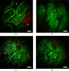
Unaveraged in vivo images of unstained rat tissue. The pseudo-color images show red SHG signal (<405 nm) and green intrinsic fluorescent emission (405-700 nm). (a) Image of the superficial kidney renal cortex shows dark renal interstitium (RI), dark cellular nuclei (N) and bright intrinsic fluorescent cytoplasm (CY) that form the epithelial cells in the renal tubules (RT), SHG signal from the tough fibrous layer that forms the renal capsule (RC), and the dark blood filled lumen (L) inside the renal tubules. (b) Image of the inner colon wall shows bright intrinsic fluorescent signal from entrocytes (E) surrounding dark circular crypts (C). (c) Image of the rat liver showing ~20 µm diameter hapatocytes (coarse dashed line) with dark nuclei (N, solid line) chained together to form hepatic chords (HC), a dark bile duct (BD, fine dashed line) and bright intrinsically fluorescent bile salts (BS), as well as SHG emission from the septa (S) a fibrous tissue bands that separates hapatocyte nodules. (d) Image of the rat liver without labels shown for clarity. In these images, scale bars are 20 µm.

Similar articles

Cited by

References

    1. Denk W., Strickler J. H., Webb W. W., “Two-photon laser scanning fluorescence microscopy,” Science 248(4951), 73–76 (1990).10.1126/science.2321027 - DOI - PubMed
    1. Lin S. J., Jee S. H., Kuo C. J., Wu R. J., Lin W. C., Chen J. S., Liao Y. H., Hsu C. J., Tsai T. F., Chen Y. F., Dong C. Y., “Discrimination of basal cell carcinoma from normal dermal stroma by quantitative multiphoton imaging,” Opt. Lett. 31(18), 2756–2758 (2006).10.1364/OL.31.002756 - DOI - PubMed
    1. Mukherjee S., Wysock J. S., Ng C. K., Akhtar M., Perner S., Lee M. M., Rubin M. A., Maxfield F. R., Webb W. W., Scherr D. S., “Human bladder cancer diagnosis using Multiphoton microscopy,” Proc. SPIE 7161, 716117–, 716117-10. (2009).10.1117/12.808314 - DOI - PMC - PubMed
    1. Pavlova I., Hume K. R., Yazinski S. A., Flanders J., Southard T. L., Weiss R. S., Webb W. W., “Multiphoton microscopy and microspectroscopy for diagnostics of inflammatory and neoplastic lung,” J. Biomed. Opt. 17(3), 036014 (2012).10.1117/1.JBO.17.3.036014 - DOI - PMC - PubMed
    1. Skala M. C., Squirrell J. M., Vrotsos K. M., Eickhoff J. C., Gendron-Fitzpatrick A., Eliceiri K. W., Ramanujam N., “Multiphoton microscopy of endogenous fluorescence differentiates normal, precancerous, and cancerous squamous epithelial tissues,” Cancer Res. 65(4), 1180–1186 (2005).10.1158/0008-5472.CAN-04-3031 - DOI - PMC - PubMed

LinkOut - more resources