. 2012 Nov;42(11):1119-30.
doi: 10.1111/j.1872-034X.2012.01026.x. Epub 2012 May 9.

Novel organotypic culture model of cholangiocarcinoma progression

Affiliations

Novel organotypic culture model of cholangiocarcinoma progression

Deanna J W Campbell et al. Hepatol Res. 2012 Nov.

Abstract

Aim: Recent studies have suggested that increased α-smooth muscle-actin positive myofibroblastic cells (α-SMA positive CAF) in the desmoplastic stroma may relate to a more aggressive cancer and worse survival outcomes for intrahepatic cholangiocarcinoma (ICC) patients. To facilitate investigating cellular and molecular interactions between α-SMA positive CAF and cholangiocarcinoma cells related to ICC progression, we developed a novel 3-D organotypic culture model of cholangiocarcinoma that more accurately mimics the stromal microenvironment, gene expression profile and select pathophysiological characteristics of desmoplastic ICC in vivo.

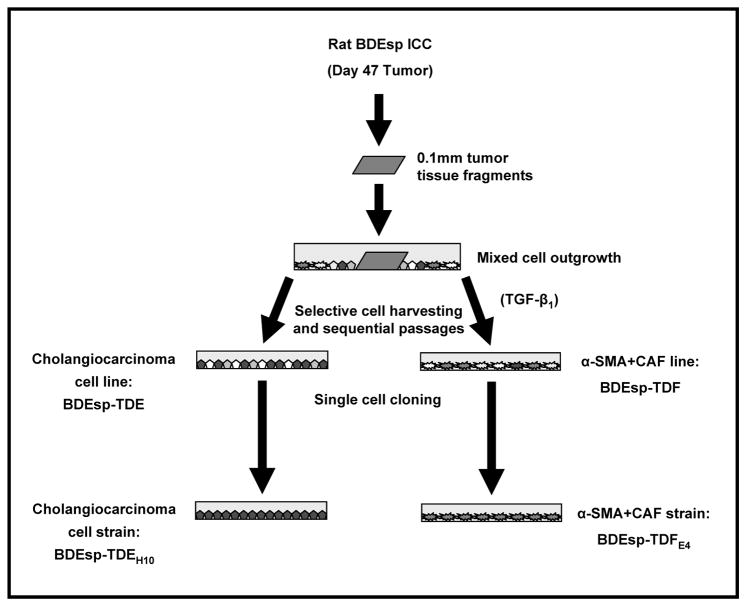
Methods: This unique model was established by co-culturing within a type I collagen gel matrix, a strain of cholangiocarcinoma cells (derived from an ICC formed in syngeneic rat liver following bile duct inoculation of spontaneously-transformed rat cholangiocytes) with varying numbers of clonal α-SMA positive CAF established from the same tumor type.

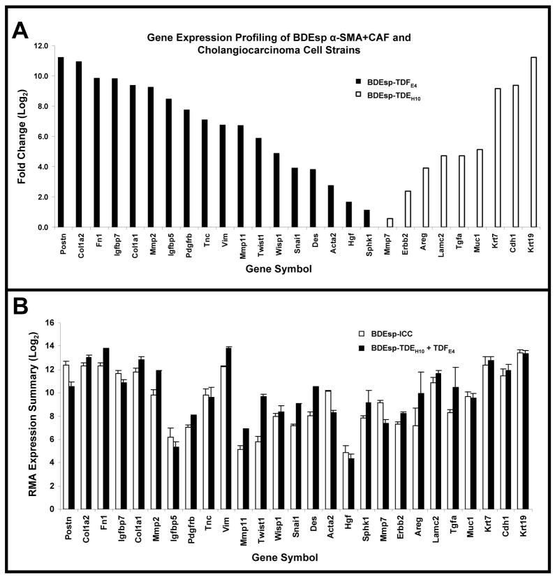
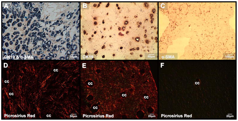
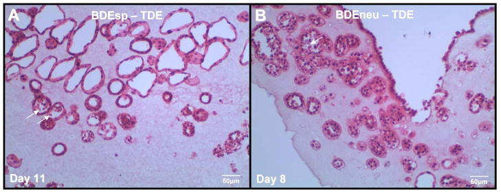
Results: Cholangiocarcinoma cells and α-SMA positive CAF in monoculture each exhibited cell-specific biomarker gene expression profiles characteristic of stromal myofibroblastic cell versus malignant cholangiocyte cell types. In comparison, the gene expression profile and histopathological characteristics exhibited by the organotypic co-culture closely resembled those of whole tissue samples of the parent orthotopic ICC. We further showed α-SMA positive CAF to significantly enhance cholangiocarcinoma cell "ductal-like" growth and cancer cell migration/invasiveness in vitro, as well as to promote upregulated expression of select genes known to be associated with ICC invasion.

Conclusion: This novel organotypic model provides an important new resource for studying the effects of microenvironment on cholangiocarcinoma progression in vitro and may have potential as a preclinical model for identifying molecularly targeted therapies.


Conflict of interest statement

Conflicts of Interest: None

Figures

Figure 1
Figure 1
Scheme outlining the method used to concomitantly establish the BDEsp-TDEH10 cholangiocarcinoma celland BDEsp-TDFE4 cancer-associated fibroblastic (CAF) cellstrains derived from an orthotopic BDEsp ICC obtained from syngeneic rat liver at day 47 after bile duct inoculation of the tumorigenic, spontaneously-transformed rat cholangiocyte cell line (BDEsp). As an initial selection step, TGF-β1 at 5ng/ml was temporarily added to the culture medium to facilitate expansion of the CAF population.
Figure 2
Figure 2
Gene expression profiling based on selected gene biomarkers of BDEsp ICC-derived cancer-associated myofibroblastic cells and cholangiocarcinoma cell strains. (A) Bar chart representation of 27 marker genes assessed by microarray analysis that showed significant differences in expression levels between BDEsp-TDFE4 and BDEsp-TDEH10 when cultured individually in a rat tail type I collagen gel matrix. (B) Robust Multiarray Analysis (RMA) expression summaries for the 27 analyzed biomarker genes shown in A, comparing orthotopic BDEsp ICC to organotypic BDEsp-TDFE4/BDEsp-TDEH10 culture. Postn, periostin; Col1a2, collagen type Ia2; Fn1, fibronectin 1; Igfbp7, insulin-like growth factor binding protein 7; Col1a1, collagen type Ia1; Mmp2, metalloproteinase-2; Igfbp5, insulin-like growth factor binding protein 5; Pdgfrb, platelet-derived growth factor receptor β; Tnc, tenascin-C; Vim, vimentin; Mmp11, metalloproteinase 11; Twist1, Twist homolog 1; Wisp1, WNT1 inducible signaling protein 1; Snail1, Snail homolog 1; Des, desmin; Acta2, smooth muscle actin; Hgf, hepatocyte growth factor; Sphk1, sphingosine kinase 1; Mmp7, metalloproteinase-7; Erbb2, c-erbB2 protooncogene receptor protein; Areg, amphiregulin; Lamc2, laminin gamma2; Tgfa, transforming growth factor α; Muc1, mucin 1; Krt7, cytokeratin 7; Cdh1, E-cadherin; Krt19, cytokeratin 19.
Figure 3
Figure 3
Co-culturing of BDEsp-TDEH10 cells with BDEsp-TDFE4 cells in rat tail type I collagen gel matrix reproduces characteristic histopathological features of desmoplastic BDEsp ICC in vivo. (A) Desmoplastic BDEsp ICC formed in rat liver showing selective positive immunostaining of well differentiated cholangiocarcinoma ducts for biliary cytokeratin 19 (CK19) (blue staining) and cancer-associated myofibroblastic cells positive for α-smooth muscle actin (α-SMA) (brown staining) in the surrounding tumor stroma. (B) Like BDEsp ICC, well differentiated “duct-like” structures formed from BDEsp-TDEH10 cells in organotypic co-culture exhibit selective immunoreactivity for CK19 (brown staining). (C) Also like BDEsp ICC, stromal BDEsp-TDFE4 cells in organotypic co-culture are selectively immunoreactive for α-SMA (brown staining). (D) Picrosirius red staining under polarized light of type I collagenous fibers (orange-yellow) densely populating the desmoplastic stroma of a BDEsp ICC. (E) Picrosirius red staining of a representative histological section of a BDEsp-TDE/BDEsp-TDF co-culture mimicking the strong extracellular staining reaction for type I collagen exhibited by the orthotopic ICC in D. (F) Picrosirius red staining of a BDEsp-TDEH10 control culture without BDEsp-TDF showing only background staining for type I collagen fibers in gel matrix. cc denotes representative cholangiocarcinoma ducts or “duct-like” structures.
Figure 4
Figure 4
Representative H & E stained histological section of our rat cholangiocarcinoma cell line BDEsp-TDE at in vitro passage 3, derived from well differentiated BDEsp ICC, and cultured alone for 11 days within rat tail type I collagen gel matrix. Note that the cholangiocarcinoma cells are organized into well differentiated “duct-like” structures reminiscent of those observed in histological sections of the parent low grade BDE ICC. For comparison, the photomicrograph in (B) demonstrates a rat cholangiocarcinoma cell line designated as BDEneu-TDE that was established in our laboratory from a more highly malignant and moderately-differentiated ICC formed in syngeneic rat liver after bile duct inoculation of oncogenic-neu transformed rat cholangiocytes. As demonstrated in B, rat BDEneu-TDE cells in organotypic culture in a type I collagen gel matrix reflect the less differentiated “duct-Like” phenotype of the parent orthotopic tumor. In both A & B, arrows point to dead cells within the lumens of some of the neoplastic “duct-like” structures, indicating that cells within the central areas of formed cholangiocarcinoma cell spheroids are likely undergoing apoptotic cell death.
Figure 5
Figure 5
Representative phase contrast photomicrographs of (A) rat BDEsp-TDEH10 cholangiocarcinoma cells cultured alone for 6 days in rat tail type I collagen gel compared with (B) in which the BDEsp-TDEH10 cells were co-cultured under comparable conditions over the same time period with BDEsp-TDFE4 CAFs. Compare with the photomicrographs of the H & E stained histological sections shown in Figure 6, noting the marked increase in 3-dimensional cell spheroids formed in the co-culture over those observed in the cholangiocarcinoma cell monoculture.
Figure 6
Figure 6
Photomicrographs of representative H & E stained histological sections of 3-dimensional organotypic cultures of BDEsp-TDEH10 cholangiocarcinomacells cultured for 6 days either in monoculture (A) or in co-culture with BDEsp-TDFE4 CAFs (B & C). Cholangiocarcinoma cells were initially plated at a cell density of 2 × 10⁵ cells/type I collagen gel in both the mono- and co-cultures. The initial CAF cell plating density in the co-cultures was at 8 × 10⁵ cells/gel. The histological section shown in B was obtained in the same experiment as that for A, whereas that in C was from a separate experiment essentially repeating the culture conditions used for B. Note the dramatic increase in the number of spheroidal/”duct-like” structures formed from the cholangiocarcinoma cells in the co-cultures (B & C) over those formed under comparable culture conditions in control cholangiocarcinoma cell monoculture without CAFs (A). Also observe a much more intense eosinophilic staining of the gel matrix in B & C compared to that of A, indicative of an increase in the cellular secretion of insoluble proteins into the co-culture matrix versus that produced in the cholangiocarcinoma cell monoculture. As further depicted in B & C, only the co-cultures exhibited prominent irregular clusters of anaplastic cholangiocarcinoma cells surrounded by stromal CAFs, reflective of malignant progression and apparent invasiveness. (D) Bar graph demonstrating the mean number of spheroidal/”duct-like” structures/cm2 tissue section area formed from BDEsp-TDEH10 cholangiocarcinoma cells to significantly increase as a function of higher initial BDEsp-TDFE4 plating densities when these ICC derived cell types were co-cultured for 6 days in rat type I collagen gel matrix. Each value represents the mean ± SD determined from microimaging measurements made on 10μm thick H & E stained histological sections (n = at least 2 sections/slide prepared from triplicate cultures for each CAF plating density). P values were determined against the 0 CAF culture condition. Gel contraction, measured as a reduction in mm gel diameter ( formula image
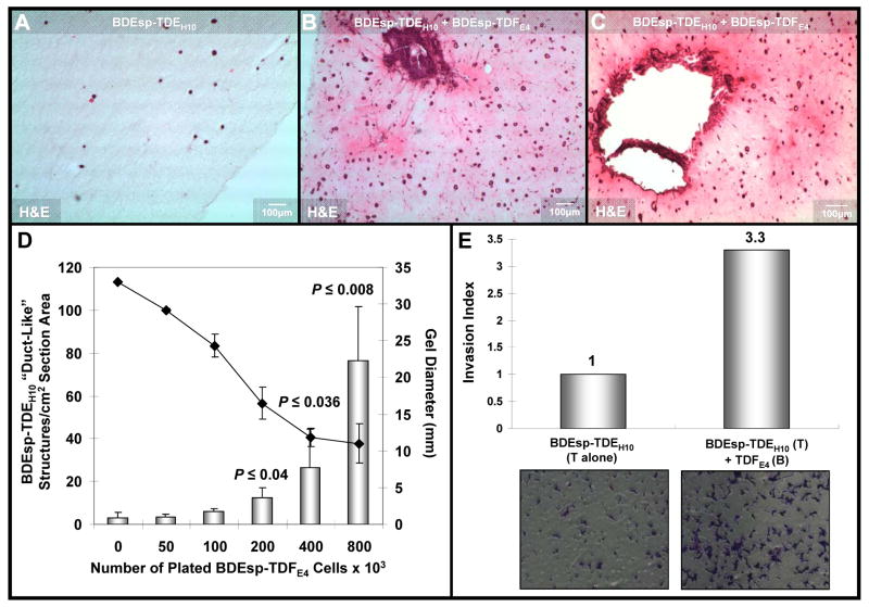
= mean ± SD) was determined to be significantly greater at the higher initial BDEsp-TDFE4 cell plating densities. (E) Representative data demonstrating BDEsp-TDFE4 CAFsto increase the cell migration/invasiveness of BDEsp-TDEH10 cholangiocarcinoma cells in vitro when assessed in the Matrigel invasion bioassay system. T= top chamber coated by Matrigel; B = bottom culture well coated with rat tail type I collagen. Invasion Index was determined according to the manufacturer’s instructions.
Figure 7
Figure 7
(A) TaqMan® QRT-PCR data demonstrating that co-culturing of rat BDEsp-TDEH10 cholangiocarcinoma cells in rat tail type I collagen gel matrix in the presence of BDEsp-TDFE4 CAFs grown to confluence on type I collagen-coated plastic significantly induced Cxcr4 gene expression in the cholangiocarcinoma cells over monoculture cell values. (B). QRT-PCR data showing conditioned medium from cultured BDEsp-TDFE4 cells to significantly up-regulate Cxcr4 gene expression in BDEsp-TDEH10 cholangiocarcinoma cells cultured alone on type I collagen-coated plastic, whereas conditioned medium from BDEsp-TDE H10 cells significantly induced Hgf gene expression in BDEsp-TDFE4 cells cultured alone under identical conditions. (C) QRT-PCR data showing a significant concentration-dependent increase in Cxcr4 gene expression in cultured BDEsp-TDEH10 cells following exposure over a 48-hour treatment period to recombinant human HGF. (D) QRT-PCR data showing the effect of conditioned medium from cultured BDEsp-TDFE4 CAFs on up-regulating Cxcr4 gene expression in cultured BDEsp-TDEH10 cholangiocarcinoma cells is partially blocked by the addition of neutralizing HGF antibody (anti-HGFab). (E) QRT-PCR data demonstrating that co-culturing of BDEsp-TDEH10 cholangiocarcinoma cells in the presence of BDEsp-TDFE4 CAFs under identical conditions as in A also produced a significant up-regulation of Muc1 gene expression in the cholangiocarcinoma cells over monoculture cell values. (F) Similar to B, conditioned medium from cultured BDEsp-TDFE4 CAFs induced a significant increase in Muc1 gene expression in cultured BDEsp-TDEH10 cholangiocarcinoma cells. PPIA (cyclophilin A) was used as the housekeeping gene to normalize Cxcr4, Hgf and Muc1 gene expression. Comparable QRT-PCR results were also obtained when normalized with Gapdh. Each value represents the mean ± SD, obtained from determinations made on a minimum of six cultures per experimental condition.

References

    1. Terada T, Makimoto K, Terayama N, et al. Alpha-smooth muscle actin-positive stromal cells in cholangiocarcinomas, hepatocellular carcinomas and metastatic liver carcinomas. J Hepatol. 1996;24:706–712. - PubMed
    1. Okamura N, Yoshida M, Shibuya A, et al. Cellular and stromal characteristics in the scirrhous hepatocellular carcinoma: comparison with hepatocellular carcinomas and intrahepatic cholangiocarcinomas. Pathol Int. 2005;55:724–731. - PubMed
    1. Sirica AE, Dumur CI, Campbell DJ, et al. Intrahepatic cholangiocarcinoma progression: prognostic factors and basic mechanisms. Clin Gastroenterol Hepatol. 2009;7:S68–S78. - PMC - PubMed
    1. Sirica AE, Campbell DJ, Dumur CI. Cancer-associated fibroblasts in intrahepatic cholangiocarcinoma. Curr Opin Gastroenterol. 2011;27:276–284. - PubMed
    1. Sirica AE. The role of cancer-associated myofibroblasts in intrahepatic cholangiocarcinoma. Nat Rev Gastroenterol Hepatol. 2012;9:44–54. - PubMed