. 2012 Jun 15;287(25):20769-73.
doi: 10.1074/jbc.C112.364620. Epub 2012 May 8.

Activation of ras signaling pathway by 8-oxoguanine DNA glycosylase bound to its excision product, 8-oxoguanine

Affiliations

Activation of ras signaling pathway by 8-oxoguanine DNA glycosylase bound to its excision product, 8-oxoguanine

Istvan Boldogh et al. J Biol Chem. .

Abstract

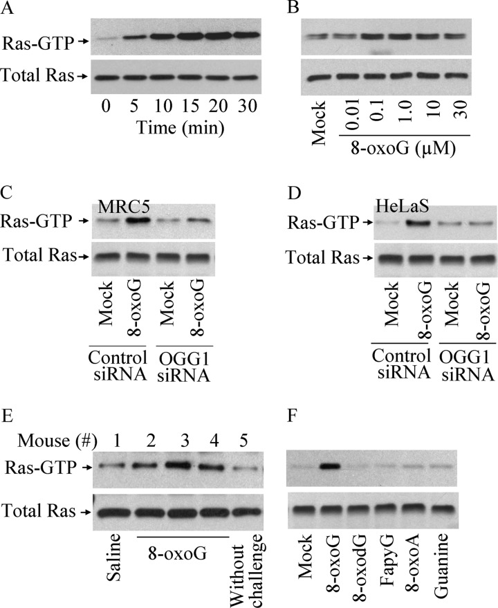
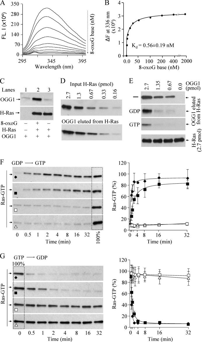
8-Oxo-7,8-dihydroguanine (8-oxoG), arguably the most abundant base lesion induced in mammalian genomes by reactive oxygen species, is repaired via the base excision repair pathway that is initiated with the excision of 8-oxoG by OGG1. Here we show that OGG1 binds the 8-oxoG base with high affinity and that the complex then interacts with canonical Ras family GTPases to catalyze replacement of GDP with GTP, thus serving as a guanine nuclear exchange factor. OGG1-mediated activation of Ras leads to phosphorylation of the mitogen-activated kinases MEK1,2/ERK1,2 and increasing downstream gene expression. These studies document for the first time that in addition to its role in repairing oxidized purines, OGG1 has an independent guanine nuclear exchange factor activity when bound to 8-oxoG.


Figures

FIGURE 1.
FIGURE 1.
Activation of Ras GTPases in OGG1-expressing cells by 8-oxoG base. A–F, parallel cell cultures were exposed to 1 μm (A, C, D, and F) or increasing concentration of 8-oxoG (B). Cell extracts were made at the times points indicated (in A) or 15 min after challenge (B, C, D, E, and F). Ras-GTP levels were determined by GST pulldown assays (15, 16). A and B, activation of Ras GTPases in a time-dependent (A) and dose-dependent manner (B) in MRC5 cells upon 8-oxoG exposure. C and D, siRNA-mediated OGG1 depletion causes decreased Ras activation. C and D, MRC5 (C) and HeLa-S (D) cells were transfected with siRNA to OGG1 (see “Experimental Procedures”) or control siRNA using two cycles of transfection and cell culture and then exposed to 8-oxoG for 15 min. Ras-GTP levels were determined as above. E, increase in Ras-GTP levels in murine lungs. BALB/c mice were challenged intranasally with 60 μl of 1 μm 8-oxoG (13), and the lungs were excised after 15 min. Ras-GTP levels in individual mouse lung extracts were determined as above. F, 8-oxoG base, but not 8-oxodG, FapyG, 8-oxoA, or guanine base increased Ras-GTP levels. 250 and 25 μg of cell extract per assay were used to determine Ras-GTP and Ras levels, respectively; n = 3–8.
FIGURE 2.
FIGURE 2.
OGG1 complexed with 8-oxoG has guanine nucleotide exchange factor activity. A, fluorescent spectroscopic analysis of 8-oxoG binding to OGG1. FL indicates fluorescence. B, the Kd was calculated from the binding isotherms shown in A (18). C, interaction of OGG1 with Ras protein requires 8-oxoG base. H-Ras (2.7 pmol) was bound to nickel-agarose beads (see “Experimental Procedures”), and OGG1 ± 8-oxoG was added. After washing three times, the amount of OGG1 bound was analyzed by immunoblotting. D, OGG1 physically interacts with Ras protein. Increasing amounts (0.16–2.7 pmol) of His-H-Ras were immobilized on nickel-agarose beads before incubation with OGG1 (2.7 pmol) plus 8-oxoG (2.7 pmol) for 1 h at 4 °C. The proteins were stripped from the beads for immunoblotting. Upper panel, His-H-Ras bound to nickel-agarose beads; lower panel, OGG1 protein eluted from H-Ras. E, guanine nucleotides decrease the OGG1 interaction with Ras. His-H-Ras (2.7 pmol) bound to agarose beads was incubated with untagged OGG1 (2.7, 1.3, 0.67, and 0 pmol) and equimolar 8-oxoG for 30 min, and a 10-fold molar excess of GTP or GDP was then added for 30 min at 24 °C. Levels of eluted proteins were determined by Western blotting. F, exchange of Ras-bound GDP for GTP. H-Ras protein (6 pmol) was loaded with GDP (6 pmol), and nucleotide exchange was initiated by adding OGG1 (6 pmol) plus 8-oxoG (♦) or 0.6 pmol of OGG1 plus 8-oxoG (■), OGG1 (□), or 8-oxoG (▴) alone, together with GTPγS (60 pmol). G, GTP-GDP exchange by OGG1. H-Ras protein (6 pmol) was loaded with GTP, and guanine nucleotide exchange was initiated by adding 6 pmol of OGG1 plus 8-oxoG (♦) or 0.6 pmol of OGG1 plus 8-oxoG (■), OGG1 (□), or 8-oxoG (▴) alone, together with 10-fold excess GDP. Ras-GTP was quantified using pulldown immunoblot assays as in Fig. 1. G and F, right panels, bands in left panels were quantitated by densitometry (ImageJ), and the time course of nucleotide exchange is depicted graphically. n = 3–4.
FIGURE 3.
FIGURE 3.
Activated Ras-mediated phosphorylation of MAPKs in 8-oxoG-treated cells. A, MRC5 cells were exposed to 1 μm 8-oxoG and lysed at the times indicated, and phosphorylated MEK 1/2 (p-MEK1/2) and -ERK1/2 (p-ERK1/2) levels were determined (upper panels) by Western blotting. Total MEK1/2 and ERK1/2 levels served as loading controls (lower panels). B, nuclear accumulation of phosphorylated ERK1/2 in MRC5 cells in response to 8-oxoG. Right panels, DAPI-stained nuclei. Scale bars: 10 μm. C, N-Ras-dependent ERK1/2 phosphorylation in MRC5 cells. Upper panel, levels of total Ras after depletion by siRNA to H-, K-, and N-Ras, shown by Western blotting. siRNA-transfected cells were treated with 1 μm 8-oxoG for 15 min, and phosphorylated ERK1/2 and ERK1/2 levels were assessed by Western blot analysis (middle and lower panels). D, isotype-specific activation of Ras in 8-oxoG-exposed MRC5 cells. Upper panel, activation of Ras isoforms after 8-oxoG addition (15 min). Ras-GTP levels were determined in 250 μg of cell extracts by pulldown assay (15, 16). Middle panel, H-, K-, and N-Ras levels in mock- and 8-oxoG-treated cells. Lower panel, total Ras in cell extracts (25 μg/lane) is shown by pan-Ras antibody. n = 3–6.

References

    1. Mitra S., Hazra T. K., Roy R., Ikeda S., Biswas T., Lock J., Boldogh I., Izumi T. (1997) Complexities of DNA base excision repair in mammalian cells. Mol. Cells 7, 305–312 - PubMed
    1. Shibutani S., Takeshita M., Grollman A. P. (1991) Insertion of specific bases during DNA synthesis past the oxidation-damaged base 8-oxodG. Nature 349, 431–434 - PubMed
    1. Cheng K. C., Cahill D. S., Kasai H., Nishimura S., Loeb L. A. (1992) 8-Hydroxyguanine, an abundant form of oxidative DNA damage, causes G → T and A → C substitutions. J. Biol. Chem. 267, 166–172 - PubMed
    1. Maga G., Villani G., Crespan E., Wimmer U., Ferrari E., Bertocci B., Hübscher U. (2007) 8-Oxo-guanine bypass by human DNA polymerases in the presence of auxiliary proteins. Nature 447, 606–608 - PubMed
    1. Haracska L., Yu S. L., Johnson R. E., Prakash L., Prakash S. (2000) Efficient and accurate replication in the presence of 7,8-dihydro-8-oxoguanine by DNA polymerase η. Nat. Genet. 25, 458–461 - PubMed

Publication types

MeSH terms