. 2012 May 8:7:68.
doi: 10.1186/1748-717X-7-68.

An athymic rat model of cutaneous radiation injury designed to study human tissue-based wound therapy

Affiliations

An athymic rat model of cutaneous radiation injury designed to study human tissue-based wound therapy

Lucas H Rifkin et al. Radiat Oncol. .

Abstract

Purpose: To describe a pilot study for a novel preclinical model used to test human tissue-based therapies in the setting of cutaneous radiation injury.

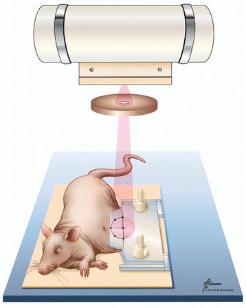
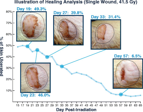
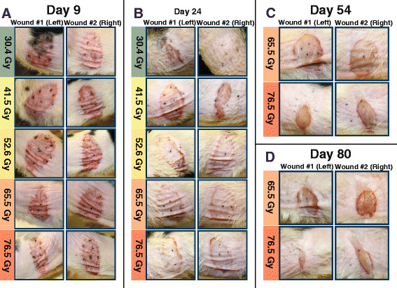
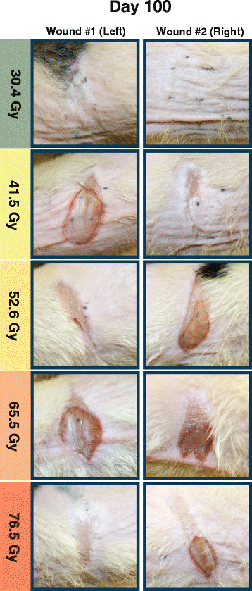
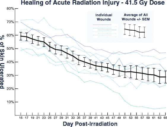
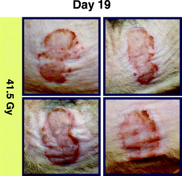
Methods: A protocol was designed to irradiate the skin of athymic rats while sparing the body and internal organs by utilizing a non-occlusive skin clamp along with an x-ray image guided stereotactic irradiator. Each rat was irradiated both on the right and the left flank with a circular field at a 20 cm source-to-surface distance (SSD). Single fractions of 30.4 Gy, 41.5 Gy, 52.6 Gy, 65.5 Gy, and 76.5 Gy were applied in a dose-finding trial. Eight additional wounds were created using the 41.5 Gy dose level. Each wound was photographed and the percentage of the irradiated area ulcerated at given time points was analyzed using ImageJ software.

Results: No systemic or lethal sequelae occurred in any animals, and all irradiated skin areas in the multi-dose trial underwent ulceration. Greater than 60% of skin within each irradiated zone underwent ulceration within ten days, with peak ulceration ranging from 62.1% to 79.8%. Peak ulceration showed a weak correlation with radiation dose (r = 0.664). Mean ulceration rate over the study period is more closely correlated to dose (r = 0.753). With the highest dose excluded due to contraction-related distortions, correlation between dose and average ulceration showed a stronger relationship (r = 0.895). Eight additional wounds created using 41.5 Gy all reached peak ulceration above 50%, with all healing significantly but incompletely by the 65-day endpoint.

Conclusions: We developed a functional preclinical model which is currently used to evaluate human tissue-based therapies in the setting of cutaneous radiation injury. Similar models may be widely applicable and useful the development of novel therapies which may improve radiotherapy management over a broad clinical spectrum.


Figures

Figure 1
Figure 1
Schematic of rat irradiation. Under anesthesia, skin was held outward in an acrylic clamp and exposed to x-ray radiation. The beam was centered on the skin edge, traversing a semicircular skin fold. As a result, a circular area of skin was irradiated.
Figure 2
Figure 2
An example of one rat wound created using a 41.5 Gy dose. The selected images show the ulceration region within the area of irradiated skin. ImageJ software was used to calculate the ulceration percentage. All data in this figure represent the same wound at different time points.
Figure 3
Figure 3
The appearance of irradiated areas throughout the 80-day period. The photographs are for five rats, one rat for each dose level. Each rat was wounded twice, once on the left side and once on the right side using the same dose. A. By day 9, erythema predominated within all irradiated areas. Crusting and desquamation were beginning to develop. Reactions intensified quickly, yet displayed a relatively uniform wound appearance across the entire dose range. Areas of increased reactivity were visible surrounding tattoo marks. B. By day 24, lower dose wounds (30.4 Gy and 41.5 Gy) each had a distinctly different appearance, while higher dose wounds (52.6 Gy to 76.5 Gy) exhibited features similar to other higher dose wounds. A 30.4 Gy wound had healed completely by day 20, accompanied by the return of hair growth in the irradiated area. Both 41.5 Gy wounds had undergone dry desquamation. Higher dose wounds displayed varying degrees of moist desquamation surrounded by intense crusting at the borders. C. By day 54, contraction in the 76.5 Gy wounds has significantly reduced the overall area of irradiated skin as well as the enclosed area of ulceration. A relatively low level of contraction was seen in the 65.5 Gy wounds. D. By day 80, contraction is apparent in the 65.5 Gy wounds, but impacts area and shape to a lesser degree than the higher dose. In the 76.5 Gy rat, contraction has distorted the irradiated area to such an extent that it has little resemblance to its original circular shape.
Figure 4
Figure 4
Photographs obtained on the final day of the study for five rats, one rat for each dose level. Significant contraction is visible in all wounds created using doses of 52.6 Gy and higher. Contraction is seen to a lesser degree on one wound (left) created using the 41.5 Gy dose. Hair growth has resumed within areas irradiated using a 30.4 Gy dose.
Figure 5
Figure 5
Healing progression of eight rats, each with one wound created using a 41.5 Gy dose. By day 65 post-irradiation, all wounds healed significantly but ulceration remained within 5.8% to 47.0% of each irradiated zone.
Figure 6
Figure 6
Four rats’ wounds in the additional group irradiated with 41.5 Gy. Appearance was similar to 41.5 Gy wounds in the multi-dose trial, without tattooing. Borders were irregular rather than maintaining a smooth, circular shape. Scabbing, crusting, and signs of healing were observed throughout wounds.

References

    1. Jemal A, Siegel R, Ward E, Murray T, Xu J, Thun MJ. Cancer Statistics, 2007. CA Cancer J Clin. 2007;57:43–66. doi: 10.3322/canjclin.57.1.43. - DOI - PubMed
    1. Jagsi R, Ben-David MA, Moran JM, Marsh RB, Griffith KA, Hayman JA, Pierce LJ. Unacceptable Cosmesis in a Protocol Investigating Intensity-Modulated Radiotherapy With Active Breathing Control for Accelerated Partial-Breast Irradiation. International Journal of Radiation Oncology Biology Physics. 2010;76:71–78. doi: 10.1016/j.ijrobp.2009.01.041. - DOI - PMC - PubMed
    1. Hopewell JW. The Skin: Its Structure and Response to Ionizing Radiation. International Journal of Radiation Biology. 1990;57:751–773. doi: 10.1080/09553009014550911. - DOI - PubMed
    1. Emami B, Lyman J, Brown A, Coia L, Goitein M, Munzenrider JE, Shank B, Solin LJ, Wesson M. Tolerance of Normal Tissue to Therapeutic Irradiation. International Journal of Radiation Oncology Biology Physics. 1991;21:109–122. - PubMed
    1. Withers HR. The Dose-survival Relationship for Irradiation of Epithelial Cells of Mouse Skin. Br J Radiol. 1967;40:187–194. doi: 10.1259/0007-1285-40-471-187. - DOI - PubMed