Review
. 2012 Jul;47(7):483-90.
doi: 10.1016/j.exger.2012.03.020. Epub 2012 Apr 28.

Aging and dry eye disease

Affiliations
Review

Aging and dry eye disease

Juan Ding et al. Exp Gerontol. 2012 Jul.

Abstract

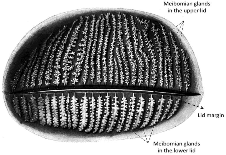
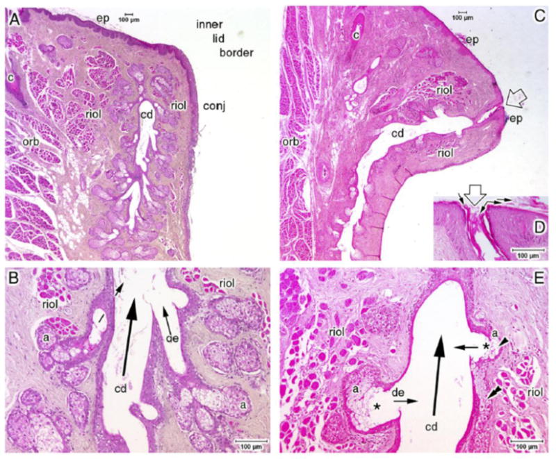
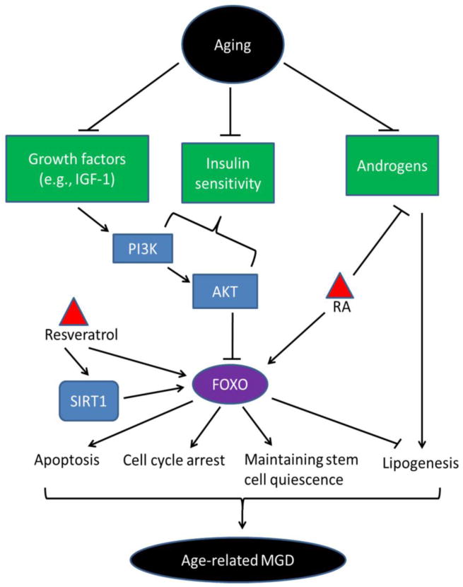
Dry eye disease is a prevalent eye disorder that in particular affects the elderly population. One of the major causes of dry eye, meibomian gland dysfunction (MGD), shows increased prevalence with aging. MGD is caused by hyperkeratinization of the ductal epithelium of meibomian gland and reduced quantity and/or quality of meibum, the holocrine product that stabilizes and prevents the evaporation of the tear film. Of note, retinoids which are used in current anti-aging cosmetics may promote the development of MGD and dry eye disease. In this review, we will discuss the possible mechanisms of age-related MGD.


Conflict of interest statement

Conflict of interest: The authors have no conflict of interest

Figures

Figure 1
Figure 1
Topography of the meibomian glands in the upper and lower eye lids. The drawing depicts a posterior view with the anterior part of the lid removed and the connective tissue made translucent so that the glands are exposed. Reproduced from Springer Science+Business Media: DER OPHTHALMOLOGE, Teil I: Anatomie, Embryologie und Histologie der Meibom-Drüsen, volume 106, 2009, page 872-983, Knop N, Knop E, Figure 3, which originated from Sobotta: Atlas der Anatomie des Menschen © Elsevier GmbH, Urban & Fischer Verlag München; with kind permission from Elsevier and Springer.
Figure 2
Figure 2
Structural comparison of the meibomian gland in normal state and obstructive MGD. (A) A normal meibomian gland. This section is not cut through the orifice of the central duct (cd). (B) is a magnification of (A) showing the central duct (cd), the connecting ductules (de) and the acini (a). Normally, the ductule (de) is narrow and enters the central duct in an oblique direction. (C–E) Histology section of a meibomian gland with obstructive MGD. (C) The orifice is indicated by the open arrow. The central duct (cd) is partly dilated, with a thinner epithelium wall than in the normal gland. (D) The orifice is obstructed by keratin lamellae which are indicated by small arrows. (E) The ductules (de) are dilated and enter the central duct (cd) at right angles (small arrows). The secretory acini (a) are smaller than in a normal gland, the number of secretory meibocytes is reduced and only a few cell layers remain (arrowhead). Other abnormal structures include: asterisks indicate formations of lumens within the acini, and the double arrowhead indicates the apparent integration of meibocytes of a disrupted acinus into the wall of the central duct. Abbreviations: a, acini; c, ciliary; cd, central duct; conj, conjunctiva mucosa; de, connecting ductules; ep, epidermis; orb, orbicularis muscle; roil, Riolan's muscle. Light microscopic images of paraffin-embedded sections stained with hematoxylin and eosin (H&E); scale bars are shown in the images. Reprinted from Knop E, Knop N, Brewitt H et al. [Meibomian glands, Part III: meibomian gland dysfunction (MGD)—plaidoyer for a discrete disease entity and as an important cause of dry eye.] Meibom-Dru¨sen, Teil III: Meibomdru¨sen Dysfunktionen (MGD)—Pla¨doyer fu¨r ein igensta¨ndiges Krankheitsbild und wichtige Ursache fu¨r das Trockene Auge. Ophthalmologe. 2009;106:966–979, Figure 5, with kind permission of Springer Science and Business Media. The legend is adapted from (Knop and others 2011).
Figure 3
Figure 3
Hypothetical signaling pathways in meibomian gland cells during aging. Levels of growth factors such as IGF-1 and androgens decline during aging, as well as insulin sensitivity, resulting in cell cycle arrest, apoptosis and reduced lipogenesis, possibly mediated via FOXOs. Increased FOXOs at old age may also keep stem cells in a quiescent state, resulting in less cell turnover of meibomian gland cells. Anti-aging agents such as RA and resveratrol also may influence FOXOs, leading to impaired meibomian gland function.

References

    1. The definition and classification of dry eye disease: report of the Definition and Classification Subcommittee of the International Dry Eye WorkShop (2007) The ocular surface. 2007;5:75–92. - PubMed
    1. Arita R, Itoh K, Inoue K, Amano S. Noncontact infrared meibography to document age-related changes of the meibomian glands in a normal population. Ophthalmology. 2008;115:911–915. - PubMed
    1. Armoni M, Harel C, Karni S, Chen H, Bar-Yoseph F, Ver MR, Quon MJ, Karnieli E. FOXO1 epresses peroxisome proliferator-activated receptor-gamma1 and -gamma2 gene promoters in primary adipocytes. A novel paradigm to increase insulin sensitivity. The Journal of biological hemistry. 2006;281:19881–19891. - PubMed
    1. Bartke A. Growth hormone, insulin and aging: the benefits of endocrine defects. Experimental gerontology. 2011;46:108–111. - PMC - PubMed
    1. Baur JA, Sinclair DA. Therapeutic potential of resveratrol: the in vivo evidence. Nature reviews Drug discovery. 2006;5:493–506. - PubMed

Publication types