Review
. 2012 Jun;139(11):1895-902.
doi: 10.1242/dev.070771.

Tet family proteins and 5-hydroxymethylcytosine in development and disease

Affiliations
Review

Tet family proteins and 5-hydroxymethylcytosine in development and disease

Li Tan et al. Development. 2012 Jun.

Abstract

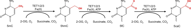
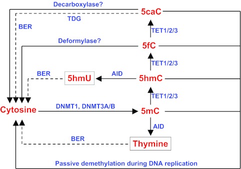
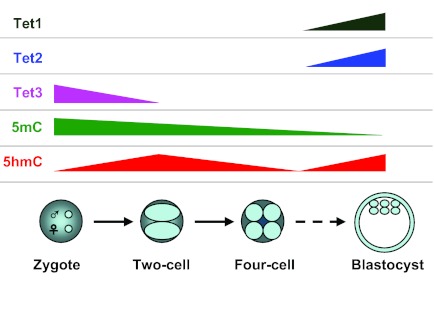
Over the past few decades, DNA methylation at the 5-position of cytosine (5-methylcytosine, 5mC) has emerged as an important epigenetic modification that plays essential roles in development, aging and disease. However, the mechanisms controlling 5mC dynamics remain elusive. Recent studies have shown that ten-eleven translocation (Tet) proteins can catalyze 5mC oxidation and generate 5mC derivatives, including 5-hydroxymethylcytosine (5hmC). The exciting discovery of these novel 5mC derivatives has begun to shed light on the dynamic nature of 5mC, and emerging evidence has shown that Tet family proteins and 5hmC are involved in normal development as well as in many diseases. In this Primer we provide an overview of the role of Tet family proteins and 5hmC in development and cancer.


Figures

Fig. 1.
Fig. 1.
Structure and function of mouse Tet family proteins. The mouse Tet protein family contains three members: Tet1, Tet2 and Tet3. All have a C-terminal CD domain (containing the Cys-rich and DSBH regions) that exhibits 2-oxoglutarate (2-OG)- and iron (II)-dependent dioxygenase activity. The CD domain also includes a spacer region, the length of which varies between Tet family members. The N-termini of Tet1 and Tet3, but not Tet2, contain a CXXC domain, which mediates their direct DNA-binding ability (Zhang et al., 2010; Xu, Y. et al., 2011). There is also a unique PRK12323 (DNA polymerase III subunits gamma and tau; provisional) domain in Tet3. This PRK12323 domain is not assigned to any domain superfamily and its function remains unknown. a.a., amino acids.
Fig. 2.
Fig. 2.
Oxidative reactions carried out by Tet proteins. Tet family proteins oxidize 5mC into 5hmC (and to 5fC and 5caC at low levels) in the presence of 2-OG and iron. 5mC and 5hmC can be further oxidized mainly to 5caC by Tet proteins if the reaction system is supplemented with ATP. 5mC, 5-methylcytosine; 5hmC, 5-hydroxymethylcytosine; 5fC, 5-formylcytosine; 5caC, 5-carboxylcytosine.
Fig. 3.
Fig. 3.
Involvement of Tet family proteins in the regulation of DNA methylation and demethylation: potential mechanisms. Tet family proteins (TET1/2/3) catalyze 5mC oxidation to 5hmC. Tet family proteins can also convert 5mC or 5hmC further into 5fC and 5caC, and the latter may be recognized and excised by thymine DNA glycosylase (TDG) to generate cytosine and thereby complete demethylation. Alternatively, because 5hmC is more sensitive than 5mC to deamination (via activation-induced deaminase, AID), it can be converted to 5-hydroxymethyluracil (5hmU), which in turn can be converted to cytosine following base-excision repair (BER) pathway-mediated demethylation (Guo et al., 2011). It remains unknown whether there are decarboxylases or deformylases that can remove the modification directly. Importantly, 5mC and its oxidative derivatives may undergo passive demethylation by dilution during DNA replication (Inoue and Zhang, 2011; Inoue et al., 2011).
Fig. 4.
Fig. 4.
Tet gene expression and 5mC and 5hmC levels during mouse preimplantation development. The expression levels of Tet1 and Tet2 are very low in oocytes and fertilized zygotes. By contrast, Tet3 is highly expressed in oocytes and fertilized zygotes but disappears rapidly during cleavage (Gu et al., 2011; Iqbal et al., 2011; Wossidlo et al., 2011). Tet3-mediated 5mC oxidation and subsequent dilution by DNA replication during embryo cleavage might contribute to the previous ‘active’ DNA demethylation observed in the developing preimplantation embryo (Gu et al., 2011; Inoue and Zhang, 2011). The expression levels of Tet1 and Tet2 increase during preimplantation development and both are highly expressed in the inner cell mass of the blastocyst, which also exhibits high 5hmC levels (Ruzov et al., 2011).

References

    1. Abdel-Wahab O., Mullally A., Hedvat C., Garcia-Manero G., Patel J., Wadleigh M., Malinge S., Yao J., Kilpivaara O., Bhat R., et al. (2009). Genetic characterization of TET1, TET2, and TET3 alterations in myeloid malignancies. Blood 114, 144–147 - PMC - PubMed
    1. Albano F., Anelli L., Zagaria A., Coccaro N., Minervini A., Rossi A. R., Specchia G. (2011). Decreased TET2 gene expression during chronic myeloid leukemia progression. Leuk. Res. 35, e220–e222 - PubMed
    1. Amir R. E., Van den Veyver I. B., Wan M., Tran C. Q., Francke U., Zoghbi H. Y. (1999). Rett syndrome is caused by mutations in X-linked MECP2, encoding methyl-CpG-binding protein 2. Nat. Genet. 23, 185–188 - PubMed
    1. Bestor T., Laudano A., Mattaliano R., Ingram V. (1988). Cloning and sequencing of a cDNA encoding DNA methyltransferase of mouse cells: the carboxyl-terminal domain of the mammalian enzymes is related to bacterial restriction methyltransferases. J. Mol. Biol. 203, 971–983 - PubMed
    1. Bestor T. H. (1992). Activation of mammalian DNA methyltransferase by cleavage of a Zn binding regulatory domain. EMBO J. 11, 2611–2617 - PMC - PubMed

Publication types

MeSH terms