. 2012 Aug;23(8):1951-9.
doi: 10.1007/s10856-012-4663-x. Epub 2012 May 9.

In vivo behaviour of a biodegradable poly(trimethylene carbonate) barrier membrane: a histological study in rats

Affiliations

In vivo behaviour of a biodegradable poly(trimethylene carbonate) barrier membrane: a histological study in rats

A C Van Leeuwen et al. J Mater Sci Mater Med. 2012 Aug.

Abstract

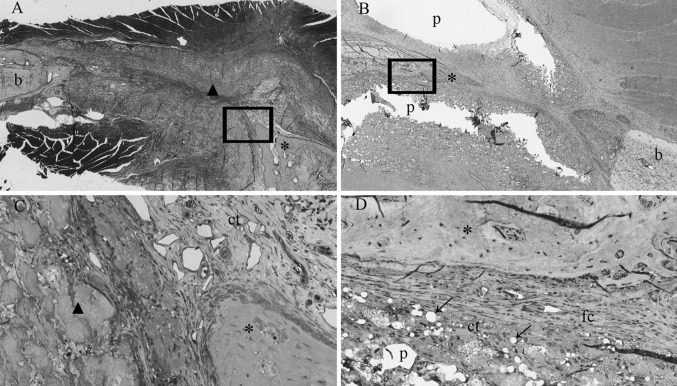
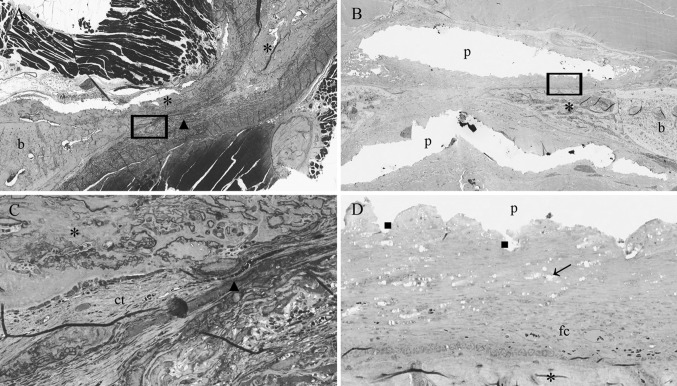
The aim of the present study was to evaluate the response of surrounding tissues to newly developed poly(trimethylene carbonate) (PTMC) membranes. Furthermore, the tissue formation beneath and the space maintaining properties of the PTMC membrane were evaluated. Results were compared with a collagen membrane (Geistlich BioGide), which served as control. Single-sided standardized 5.0 mm circular bicortical defects were created in the mandibular angle of rats. Defects were covered with either the PTMC membrane or a collagen membrane. After 2, 4 and 12 weeks rats were sacrificed and histology was performed. The PTMC membranes induced a mild tissue reaction corresponding to a normal foreign body reaction. The PTMC membranes showed minimal cellular capsule formation and showed signs of a surface erosion process. Bone tissue formed beneath the PTMC membranes comparable to that beneath the collagen membranes. The space maintaining properties of the PTMC membranes were superior to those of the collagen membrane. Newly developed PTMC membranes can be used with success as barrier membranes in critical size rat mandibular defects.


Figures

Fig. 1
Fig. 1
Light micrographs of rat mandibular defects covered with either a collagen (a, c) or a PTMC (b, d) membrane after 2 weeks of implantation. The collagen and PTMC membrane can be readily distinguished. Degradation of the PTMC membrane can be observed. Toluidin blue with basic fuchsin as counterstain was used for the staining of the histological sections. (p) polymer, (ct) connective tissue, (b) bone, (fc) fibrous capsule, (asterisk) osteoid, (filled triangle) collagen membrane, arrows indicate phagocytosed intracellular fragments. Figures ‘c and d’ magnifications (×20) of the marked regions from respectively figures ‘a and b’, which are ×2 magnifications
Fig. 2
Fig. 2
Light micrographs of rat mandibular defects covered with either a collagen (a, c) or a PTMC (b, d) membrane after 4 weeks of implantation. The collagen membrane can be distinguished from the rats connective tissue. Bone forms in and around the collagen membrane, by contrats bone formation underneath the PTMC membrane originates from the defect borders. Toluidin blue with basic fuchsin as counterstain was used for the staining of the histological sections. (p) polymer, (ct) connective tissue, (b) bone, (fc) fibrous capsule, (asterisk) osteoid, (filled triangle) collagen membrane, arrows indicate phagocytosed intracellular fragments. (filled square) Indicates surface erosion degradation by macrophage and giant cell activity. Figures ‘c and d’ magnifications (×20) of the marked regions from respectively figures ‘a and b’, which are ×2 magnifications
Fig. 3
Fig. 3
Light micrographs of rat mandibular defects covered with either a collagen (a, c) or a PTMC (b, d) membrane after 12 weeks of implantation. Bone bridges the mandibular defects in both membrane groups. The collagen membrane is not present anymore and has resorbed. The PTMC membrane eroded completely, only a few phagocytosed polymer particles are in situ. Toluidin blue with basic fuchsin as counterstain was used for the staining of the histological sections. (p) polymer, (ct) connective tissue, (b) bone, (fc) fibrous capsule, (asterisk) osteoid, (v) marks blood vessel, (f) fat cell, arrows indicate phagocytosed intracellular fragments. Figures ‘c and d’ are magnifications (×20) of the marked regions from respectively figures ‘a and b’, which are ×2 magnifications
Fig. 4
Fig. 4
a Histological scoring results for the soft tissue response to the collagen and PTMC membrane after 2, 4 and 12 weeks. Error bars represent means ± standard deviation for n = 4, *p < 0.05. b Scoring results for the proliferated tissue at the mandibular defect site. Error bars represent means ± standard deviation for n = 4, *p < 0.05. c Scoring results for the space-maintaining properties for both membranes. At time = 12 weeks the space-maintaining properties could not be assessed because of closure of the mandibular defects and degradation of both the membranes. Error bars represent means ± standard deviation for n = 4, *p < 0.05

References

    1. Hollinger JO, Buck DC, Bruder SP. Biology of bone healing: its impact on clinical therapy. In: Lynch SE, Genco RJ, Marx RE, editors. Tissue engineering. Applications in maxillofacial surgery and periodontics. Chicago: Quintessence; 1999. pp. 17–53.
    1. Kay SA, Wisner-Lynch L, Marxer M, Lynch SE. Guided bone regeneration: integration of a resorbable membrane and a bone graft material. Practical periodontics and aesthetic dentistry. PPAD. 1997;9:185–196. - PubMed
    1. von Arx T, Cochran DL, Schenk RK, Buser D. Evaluation of a prototype trilayer membrane (PTLM) for lateral ridge augmentation: an experimental study in the canine mandible. Int J Oral Maxillofac Surg. 2002;31:190–199. doi: 10.1054/ijom.2001.0205. - DOI - PubMed
    1. Pauli G. Tissue safety in view of CJD and variant CJD. Cell Tissue Bank. 2005;6:191–200. doi: 10.1007/s10561-005-0336-z. - DOI - PubMed
    1. Fishman JA. Infection in xenotransplantation. J Card Surg. 2001;16:363–373. doi: 10.1111/j.1540-8191.2001.tb00536.x. - DOI - PubMed

LinkOut - more resources