Comparative Study
. 2012 Jun;122(6):1317-25.
doi: 10.1002/lary.23307. Epub 2012 May 8.

Pepsin promotes proliferation of laryngeal and pharyngeal epithelial cells

Affiliations
Comparative Study

Pepsin promotes proliferation of laryngeal and pharyngeal epithelial cells

Nikki Johnston et al. Laryngoscope. 2012 Jun.

Abstract

Objective/hypothesis: Laryngopharyngeal reflux (LPR) is thought to be a significant risk factor for laryngeal squamous cell carcinoma (SCC), but causality has never been proven. It is accepted that chronic reflux into the esophagus can induce metaplastic changes in esophageal mucosa with subsequent increased risk of esophageal adenocarcinoma, but no similar associations have been established for LPR and laryngopharyngeal SCC. The objective of this study was to test the hypothesis that reflux of pepsin into the laryngopharynx can promote carcinogenesis.

Study design: Translational research study.

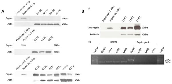
Methods: Normal human laryngeal primary epithelial cell cultures and hypopharyngeal FaDu SCC cells were exposed to human pepsin and analyzed by Human Cancer PathwayFinder and miRNA Superarrays, flow cytometry, and Western blot to determine the effect of pepsin on carcinogenesis. Laryngeal biopsy specimens taken from cancer patients and normal control subjects were analyzed for the presence of pepsin by Western blot.

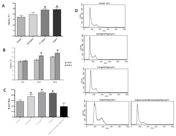
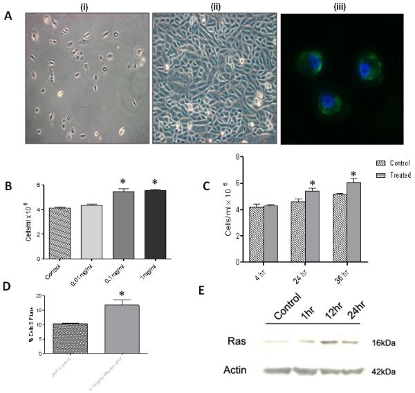
Results: Microarray analysis demonstrated that pepsin significantly altered the expression of 27 genes implicated in carcinogenesis and also affected the expression of 22 microRNAs known to be altered in human head and neck cancers. Pepsin increased proliferation in both FaDu SCC cells and cultured normal laryngeal epithelial primary cells by increasing S phase distribution on flow cytometry analysis in a time- and dose-dependent manner. Furthermore, pepsin was detected in 60% (3/5) human laryngeal cancer biopsies, absent in all (0/5) normal control specimens.

Conclusions: These data support a role for refluxed pepsin in the promotion of epithelial proliferation and carcinogenesis of the larynx and pharynx.


Figures

Figure 1
Figure 1. Pepsin increases growth and proliferation of FaDu SCC cells
(A) FaDu cells exposed to pepsin at pH7 for 1 hour at 37C, washed 3 × briefly and incubated in fresh media for 24 hours at 37C. Cells stained with trypan blue and counted using a hemocytometer. Pepsin exposure (0.1mg/ml and 1mg/ml) results in a significant dose response increase in cell number (p<0.01). (B) Cells exposed to 0.1mg/ml pepsin at pH7 for 1 hour at 37C, washed 3 × briefly and incubated in fresh media for 4, 24, or 36 hours as indicated. A significant time response increase (24hr and 36hr) in cell number is observed in cells exposed to pepsin (p<0.001). Data from 5 biological replicates for dose response experiments were analyzed by one-way analysis of variance and Tukey’s multiple comparisons post test. Data from 5 biological replicates for time response experiments were analyzed by two-way analysis of variance and the Bonferroni multiple comparisons post test. Bar graphs show mean and standard deviation. (C) FaDu cells exposed to pepsin at pH7 for 1 hour at 37C, washed 3 × briefly, and incubated in fresh media for 24 hours at 37C. Cells were fixed, stained with propidium iodide and analyzed by flow cytometry. Data from 5 biological replicates were analyzed by one-way analysis of variance and Tukey’s multiple comparisons post test. Bar chart shows mean and standard deviation. There is a dose response increase in % cells in S phase when treated with 0.01mg/ml pepsin relative to control (p<0.05), with a further increase in % cells in S phase when exposed to pepsin at 0.1 and 1mg/ml (p<0.001). An increase in % cells in S phase is not seen when cells are treated with irreversibly inactivated pepsin. (D) Representative histograms from cell cycle analysis.
Figure 2
Figure 2. Pepsin increases growth and proliferation of normal laryngeal primary epithelial cells
(A) Isolation and culture of primary human laryngeal epithelial cells from normal biopsy specimens. Characterization as in Rees et al. using light microscopy and immunohistochemistry. i) Phase contrast microscopy of primary laryngeal epithelial cells at 5 days, 10× magnification. ii) 15 days, 20× magnification. Characteristic epithelioid shape showing a pavement-like arrangement. iii) The epithelial phenotype of cells was also confirmed by expression of cytokeratins 5 and 8 (green fluorescence) on cell cytospins by immunofluorescence. Cell nuclei were stained with DAPI (blue fluorescence), 40× magnification (45). (B) Primary epithelial laryngeal cells exposed to pepsin at pH7 for 1 hour at 37C, washed 3 × briefly and incubated in fresh media for (i) 24 hours at 37C. Cells stained with trypan blue and counted using a hemocytometer. Pepsin exposure (0.1mg/ml and 1mg/ml) results in a significant dose response increase in cell number (p<0.01). (C) Cells exposed to 0.1mg/ml pepsin at pH7 for 1 hour at 37C, washed 3 × briefly and incubated in fresh media for 4, 24, or 36 hours as indicated. A significant time response increase (24hr and 36hr) in cell number is observed in cells exposed to pepsin (p<0.001). (D) Click-iT EdU proliferation assay. Mean ± SEM % cells in S phase. Pepsin (0.1mg/ml at pH7 for 1 hour, washed 3 times and incubated in growth media for 24 hours at 37C) significantly increases proliferation of primary epithelial cells (p<0.001). (E) Western blot analysis for Ras protein levels in normal human laryngeal epithelial primary cells exposed to pepsin (0.1mg/ml, pH7) for 1, 12 and 24 hours at 37C and pH7 control. Ras protein levels increased following exposure to pepsin.
Figure 3
Figure 3. Pepsin is detected in human laryngeal cancer biopsies, absent in normal laryngeal control specimens
(A) Western blot analysis for pepsin in human tissue biopsy specimens. Pepsin was detected in 3/5 cancer patients (C1-C3), but not in any of the normal control subjects (0/5; N1-N5). SG = subglottic, PC = posterior cricoids, Tr – trachea, Tu = tumor. 30ug total protein was loaded for all except C1PC (0.6ug), C2PC (1.5ug) and C2Tu (10ug). (B) Presence of pepsin in LPR patient laryngeal biopsies and corresponding absence of pepsin precursor mRNA. (i) Pepsin was detected in all samples. To verify that pepsin observed by western blot was not synthesized locally, a section of the biopsy was analyzed for presence of pepsinogen mRNA via reverse transcriptase polymerase chain reaction (RT-PCR) (ii). Pepsinogen mRNA control testing was negative. Amplicon corresponding to pepsinogen A (437bp, expected size) was detected in human gastric tissue, but not in laryngeal specimens. HPRT1 was detected in all samples (307bp, expected size).

References

    1. Berrino F. Survival of cancer patients in Finland, 1955-1994. Acta oncologica (Stockholm, Sweden) 1999;38:275–277. - PubMed
    1. Jemal A, Murray T, Ward E, et al. Cancer statistics, 2005. CA: a cancer journal for clinicians. 2005;55:10–30. - PubMed
    1. Rothman KJ, Cann CI, Flanders D, Fried MP. Epidemiology of laryngeal cancer. Epidemiologic reviews. 1980;2:195–209. - PubMed
    1. Koufman JA. The otolaryngologic manifestations of gastroesophageal reflux disease (GERD): a clinical investigation of 225 patients using ambulatory 24-hour pH monitoring and an experimental investigation of the role of acid and pepsin in the development of laryngeal injury. The Laryngoscope. 1991;101:1–78. - PubMed
    1. Copper MP, Smit CF, Stanojcic LD, Devriese PP, Schouwenburg PF, Mathus-Vliegen LM. High incidence of laryngopharyngeal reflux in patients with head and neck cancer. The Laryngoscope. 2000;110:1007–1011. - PubMed

Publication types

MeSH terms