. 2012 Jun 5;125(22):2751-61.
doi: 10.1161/CIRCULATIONAHA.111.044354. Epub 2012 May 8.

Targeted deletion of microRNA-22 promotes stress-induced cardiac dilation and contractile dysfunction

Affiliations

Targeted deletion of microRNA-22 promotes stress-induced cardiac dilation and contractile dysfunction

Priyatansh Gurha et al. Circulation. .

Abstract

Background: Delineating the role of microRNAs (miRNAs) in the posttranscriptional gene regulation offers new insights into how the heart adapts to pathological stress. We developed a knockout of miR-22 in mice and investigated its function in the heart.

Methods and results: Here, we show that miR-22-deficient mice are impaired in inotropic and lusitropic response to acute stress by dobutamine. Furthermore, the absence of miR-22 sensitized mice to cardiac decompensation and left ventricular dilation after long-term stimulation by pressure overload. Calcium transient analysis revealed reduced sarcoplasmic reticulum Ca(2+) load in association with repressed sarcoplasmic reticulum Ca(2+) ATPase activity in mutant myocytes. Genetic ablation of miR-22 also led to a decrease in cardiac expression levels for Serca2a and muscle-restricted genes encoding proteins in the vicinity of the cardiac Z disk/titin cytoskeleton. These phenotypes were attributed in part to inappropriate repression of serum response factor activity in stressed hearts. Global analysis revealed increased expression of the transcriptional/translational repressor purine-rich element binding protein B, a highly conserved miR-22 target implicated in the negative control of muscle expression.

Conclusion: These data indicate that miR-22 functions as an integrator of Ca(2+) homeostasis and myofibrillar protein content during stress in the heart and shed light on the mechanisms that enhance propensity toward heart failure.


Figures
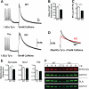
Figure 1
Figure 1
Decreased sarcoplasmic reticulum (SR) Ca2+ ATPase (SERCA2) cardiac activity and SR Ca2+ load in miR-22–/– (knockout [KO]) mice. A, Representative [Ca2+]i recordings obtained from Fluo-4 am–loaded cardiomyocytes from wild-type (WT) and KO mice during 1-Hz pacing in 1.8 mmol/L Ca2+, Tyrode solution. B through D, Rate of Ca2+ transient decay was quantitatively slower in KO mice. SR Ca2+ load was measured by changing super-fusate to 10 mmol/L caffeine in 0 Na+, 0 Ca2+ Tyrode solution, and data were analyzed with a linear mixed statistical model as described in the Methods section in the online-only Data Supplement. SR load was ascertained by quantitative ratio of fluorescent F1 to F0. Total numbers of cardiac myocytes analyzed from 3 mice of each genotype were 24 (WT) and 23 (KO). *P<0.05; ***P<0.005. E, Serca2a, Plb, and Ncx1 cardiac mRNA expression levels were detected by quantitative polymerase chain reaction from 12-week old KO and WT mice. (n=3). F, Western blot for SERCA2 and phospholamban (PLB) cardiac expression (pentameric) from pools of 3 mice of each genotype. Mann–Whitney U test: *P<0.05.
Figure 2
Figure 2
Accelerated maladaptive remodeling and cardiac dysfunction in transverse aortic constriction (TAC)–operated miR-22–/– (knockout [KO]) mice. A, Masson trichrome–stained histological sections of hearts taken from wild-type (WT) and KO mice subjected to 4 weeks of sham or TAC operation. Scale bars=1 mm (top) or 50 mm (bottom). B, Analyses of cardiac function from sham- or TAC-operated mice by echocardiography showing accelerated cardiac dysfunction and left ventricular (LV) dilation in mutant mice after 4 weeks. LVESD indicates LV end-systolic dimension; LVEDD, LV end-diastolic dimension; LVPWS, LV posterior wall thickness at end systole; and FS, fractional shortening. WT: n=5 sham and 9 TAC; KO: n=6 sham and 11 TAC. C, Quantification of picrosirius red–stained sections revealed increased fibrosis after 4 weeks in TAC-operated KO mice (n=4). D, Histological assessment of von Kossa–stained cardiac sections showed regions of calcified deposits in KO mice after 4 weeks of TAC. E, Heart weight/tibia length (HW/TL) measurements from indicated mice after 4 weeks. WT: n=5 sham and 9 TAC; KO: n=10 sham and 14 TAC. F, Myh7 and miR-208b cardiac expression was determined by quantitative polymerase chain reaction after 4 weeks of surgery (n=3–5). G, Western blot analysis revealed that beta-myosin heavy chain protein expression was blocked in KO mice subjected to 4 weeks of TAC surgery. Vertical white lines denote nonadjacent lanes on same blot. β-MHC indicates β-myosin heavy chain. Student t test (C) or 2-way ANOVA with the Tukey post hoc test (B, E, F): *P<0.05, **P<0.005, and ***P<0.0005.
Figure 3
Figure 3
Impaired sarcoplasmic reticulum (SR) Ca2+ ATPase (SERCA2) expression in transverse aortic constriction (TAC)–operated miR-22–/– (knockout [KO]) mice. A, Expression levels of indicated genes were quantified by quantitative polymerase chain reaction after 1 week of TAC (n=4), B, Representative Western blot analysis of SERCA2, calsequestrin (CASQ), monomeric phospholamban (PLB), PLB–Ser-16, and PLB–Thr-17 after 1 week of TAC. Densitometry analysis showed a reduction of these proteins after TAC (n=3–5 immunoblots from 3 mice of each genotype). Vertical white lines denote nonadjacent lanes on same blot. Student t test (B) or 2-way ANOVA with the Tukey post hoc test (A): *P<0.05, **P<0.005, and ***P<0.0005.
Figure 4
Figure 4
Global repression of myofibrillar/contractile genes in miR-22–/– (knockout [KO]) mice. A, Gene set enrichment analysis (GSEA) analysis revealed global repression of genes annotated as contractile fiber components in KO hearts (false discovery rate q value=0.00096). B through D, Gene expression was evaluated by quantitative polymerase chain reaction analysis from wild-type (WT) or miR-22–/– mice subjected to sham or transverse aortic constriction (TAC) surgery for 1 week (n=3–4). Statistical significance by 2-way ANOVA followed by the Tukey post hoc test. GO indicates gene ontology.
Figure 5
Figure 5
Increased expression of purine-rich element binding protein B (PURB) in miR-22–/– (knockout [KO]) adult hearts. A, Sylamer analysis revealed that only miR-22 seed matches were enriched among upregulated genes in KO hearts. miR-22 and murine microRNA seed matches are represented as blue and gray lines, respectively. Transcripts were sorted from upregulated to downregulated in the KO. The orange vertical line denotes the cutoff with a 5% false discovery rate (FDR). Horizontal dashed lines represent Sylamer P value thresholds of 0.01. B, Pie chart representing the distribution of upregulated transcripts containing miR-22 seed matches within the 3′ untranslated region (UTR). C, quantitative polymerase chain reaction (qPCR) detection of Purb mRNA in mice after 4 weeks of transverse aortic constriction (TAC; n=3–5). D, Western blot of PURB from indicated mice at 4 weeks of TAC. Protein levels of PURB were evaluated from the intensity of bands by densitometry. The PURB antibody used also recognizes purine-rich element binding protein A (PURA) (n=3–5 immunoblots from 3 mice of each genotype). Vertical white lines denote nonadjacent lanes on the same blot. E, Purb mRNA expression determined by qPCR from ventricular cardiomyocytes isolated by sedimentation and Percoll gradient centrifugation (n=3). F, Schematic shows that Purb contains four 3′ UTR matches to seed of miR-22. Alignments for 2 regions are shown in the indicated species. G, Wild-type (WT) or binding site mutant (mut) 3′ UTRs of Purb were cloned downstream of the renilla luciferase reporter. Plasmids were cotransfected with either a miR-22 (red bars) or control Cel-miR-64 (blue bars) duplex into cultured cells. Reporter activity was measured 24 hours after transfection and normalized to firefly activity. Data are the mean±SEM from 2 experiments carried out in triplicate. Student t test (E) or 2-way ANOVA with the Tukey post hoc test (C, D, G): *P<0.05, **P<0.005, and ***P<0.0005.
Figure 6
Figure 6
Transgenic cardiac overexpression of miR-22 induces cardiac hypertrophy. A, miR-22 expression was determined by quantitative polymerase chain reaction (qPCR) from ventricles of wild-type (WT), miR-22-Tg low (TG-low), and miR-22-Tg high (TG-high) line mice 5 weeks of age (n=3–4). B, The heart weight/tibia length (HW/TL) ratios of both miR-22 transgenic lines were significantly higher than for WT controls at 5 weeks of age (n=5–6). C and D, Wheat germ agglutinin–stained sections and cross-sectional areas (CSAs) from hearts of WT and miR-22-Tg mice (scale bars=50 mm; n=4). E through G, Cardiac mRNA expression levels were obtained by qPCR from indicated mice (n=3–4). One-way ANOVA followed by the post hoc Dunnett test: *P<0.05; **P<0.005, ***P<0.0005.
Figure 7
Figure 7
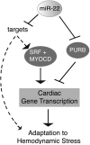
miR-22 functions to maintain muscle-restricted gene expression during pressure overload in the heart. Cardiac deletion of miR-22 accelerates the onset of maladaptation to pathological stress in part due to inappropriate repression of CArG genes involved in excitation-contraction coupling, structural integrity, and cardiac contractile force. We speculate that increased purine-rich element binding protein B (PURB) expression in the absence of miR-22 interferes with muscle-restricted gene expression directly through increased binding to repressive purine nucleotide–rich (PNR) elements found in promoters of individual genes and/or indirectly by interfering with serum response factor (SRF)–myocardin (MYOCD). miR-22 is likely to titrate the expression of numerous mRNA targets in the heart to influence other aspects of adaptation to hemodynamic stress.

Comment in

References

    1. Diwan A, Dorn GW., 2nd Decompensation of cardiac hypertrophy: cellular mechanisms and novel therapeutic targets. Physiology (Bethesda) 2007;22:56–64. - PubMed
    1. Frey N, Olson EN. Cardiac hypertrophy: the good, the bad, and the ugly. Annu Rev Physiol. 2003;65:45–79. - PubMed
    1. Periasamy M, Huke S. SERCA pump level is a critical determinant of Ca(2+)homeostasis and cardiac contractility. J Mol Cell Cardiol. 2001;33:1053–1063. - PubMed
    1. Ahmad F, Seidman JG, Seidman CE. The genetic basis for cardiac remodeling. Annu Rev Genomics Hum Genet. 2005;6:185–216. - PubMed
    1. Bers DM. Cardiac excitation-contraction coupling. Nature. 2002;415:198–205. - PubMed

Publication types

MeSH terms