. 2012 Jul 15;590(14):3305-16.
doi: 10.1113/jphysiol.2012.229690. Epub 2012 May 8.

Translational evidence that impaired autophagy contributes to arterial ageing

Affiliations

Translational evidence that impaired autophagy contributes to arterial ageing

Thomas J LaRocca et al. J Physiol. .

Abstract

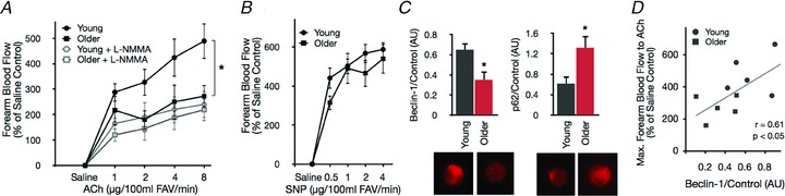
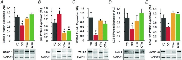
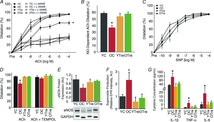
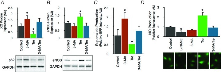
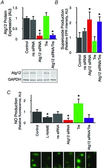
Ageing causes arterial endothelial dysfunction that increases the risk of cardiovascular diseases (CVD), but the underlying mechanisms are incompletely understood. The aim of the present study was to determine the role of autophagy, the cellular process of recycling damaged biomolecules, in endothelial dysfunction with ageing. In older humans, expression of autophagy markers in arterial endothelial cells was impaired by ∼50% (P <0.05) and was associated with an ∼30% (P <0.05) reduction in arterial endothelium-dependent dilatation (EDD). Similarly, in C57BL/6 control mice ageing was associated with an ∼40% decrease (P <0.05) in arterial markers of autophagy and an ∼25% reduction (P <0.05) in EDD. In both humans and mice, impaired EDD was mediated by reduced nitric oxide (NO) bioavailability and was associated with increased oxidative stress and inflammation (P <0.05). In old mice, treatment with the autophagy-enhancing agent trehalose restored expression of autophagy markers, rescued NO-mediated EDD by reducing oxidative stress, and normalized inflammatory cytokine expression. In cultured endothelial cells, inhibition of autophagy increased oxidative stress and reduced NO production, whereas trehalose enhanced NO production via an autophagy-dependent mechanism. These results provide the first evidence that autophagy is impaired with ageing in vascular tissues. Our findings also suggest that autophagy preserves arterial endothelial function by reducing oxidative stress and inflammation and increasing NO bioavailability. Autophagy-enhancing strategies may therefore have therapeutic efficacy for ameliorating age-associated arterial dysfunction and preventing CVD.


Figures

Figure 1
Figure 1. Impaired EDD in older humans is associated with reduced markers of autophagy
A and B, forearm blood flow responses to intra-brachial infusion of the endothelium-dependent dilator acetylcholine (ACh) in the absence and presence of the eNOS inhibitor NG-monomethyl-l-arginine (l-NMMA), and to the endothelium-independent dilator sodium nitroprusside (SNP) in young and older healthy adults. FAV, forearm volume. C, expression of the key autophagy proteins beclin 1 and p62 in endothelial cells isolated from the brachial artery. D, correlation between maximal forearm blood flow to ACh and beclin 1 protein expression. Protein expression values normalized to HUVEC control cells. All values are means ± SEM (n= 5 per group). *P < 0.05 vs. young.
Figure 2
Figure 2. Autophagy is impaired in the vasculature of old mice and restored by trehalose supplementation
A, key autophagy mediator beclin 1 in aorta of young and old control (YC and OC) and young and old trehalose supplemented (YTre and OTre) mice. B, p62, a marker of undegraded autophagy substrates. C and D, WIPI-1 and LC3-II, markers/indexes of macroautophagy. E, LAMP-2a, critical mediator of chaperone-mediated autophagy. Data expressed relative to GAPDH and normalized to YC mean value. Representative Western blot images below. Values are means ± SEM (n= 5–7 per group). *P < 0.05 vs. YC.
Figure 3
Figure 3. Impaired EDD in old mice is mediated by excessive superoxide, reduced NO bioavailability and increased inflammation, and is restored by trehalose supplementation
A, dose responses to the endothelium-dependent dilator acetylcholine (ACh) in the absence and presence of the eNOS inhibitor l-NAME in carotid arteries of young and old control (YC and OC) and young and old trehalose-supplemented (YTre and OTre) mice. B, NO-dependent dilatation (Max DilatationACh– Max DilatationACh + L-NAME). C, dose responses to the endothelium-independent dilator sodium nitroprusside (SNP). D, maximal dilatation of carotid arteries to ACh in the presence/absence of TEMPOL a superoxide dismutase mimetic. E, aortic protein expression of eNOS expressed relative to GAPDH and normalized to YC mean value. Representative Western blot image below. F, mean EPR signal for superoxide from aortic rings. G, aortic expression of the inflammatory cytokines IL-1β, TNFα, and IL-6. Values are means ± SEM (n= 5–7 per group). *P < 0.05 vs. YC.
Figure 4
Figure 4. Autophagy influences human endothelial cell function by modulating oxidative stress and NO bioavailability
A and B, protein expression of p62 and eNOS in control HUVECs and HUVECs treated for 24 h with the autophagy inhibitor 3-MA (10 mm) and/or trehalose (10 mm). Data expressed relative to GAPDH and normalized to control mean value. Representative Western blot images below. C, mean EPR signal for superoxide in control HUVECs and cells treated with 3-MA and/or trehalose. D, NO production assessed by fluorescence microscopy with the NO-specific dye DAF-FM-DA in control HUVECs and HUVECs treated with the eNOS inhibitor l-NAME, 3-MA and/or trehalose. Representative fluorescence images below. Values are means ± SEM. *P < 0.05 vs. control.
Figure 5
Figure 5. Autophagy is directly linked with NO homeostasis and the effects of trehalose
A, Atg12 protein expression in control HUVECs and HUVECs transfected with nonsense (ns) or Atg12-specific siRNA and/or treated with trehalose. Representative Western blot image below. B, mean EPR signal for superoxide in control HUVECs and cells treated with siRNA and/or trehalose. C, NO production assessed with DAF-FM-DA in control HUVECs and cells treated with siRNA and/or trehalose. Representative fluorescence images below. Values are means ± SEM. *P < 0.05 vs. control.
Figure 6
Figure 6. Autophagic flux is reduced in vascular tissue of old mice and enhanced by trehalose
Top, representative Western blot images of p62 protein in aorta from young and old control mice (YC and OC). Isolated aortas were incubated at 37°C with or without trehalose (Tre, 10 mm), and segments of aorta were snap frozen at the indicated time points. Bottom, autophagic protein degradation expressed as change in p62 levels relative to β-actin. Values are means ± SEM (n= 4 per group). *P < 0.05 vs. YC.

References

    1. Arai C, Arai N, Mizote A, Kohno K, Iwaki K, Hanaya T, Arai S, Ushio S, Fukuda S. Trehalose prevents adipocyte hypertrophy and mitigates insulin resistance. Nutr Res. 2010;30:840–848. - PubMed
    1. Bjorkoy G, Lamark T, Pankiv S, Overvatn A, Brech A, Johansen T. Monitoring autophagic degradation of p62/SQSTM1. Methods Enzymol. 2009;452:181–197. - PubMed
    1. Blackwell KA, Sorenson JP, Richardson DM, Smith LA, Suda O, Nath K, Katusic ZS. Mechanisms of aging-induced impairment of endothelium-dependent relaxation: role of tetrahydrobiopterin. Am J Physiol Heart Circ Physiol. 2004;287:H2448–2453. - PubMed
    1. Bouis D, Hospers GA, Meijer C, Molema G, Mulder NH. Endothelium in vitro: a review of human vascular endothelial cell lines for blood vessel-related research. Angiogenesis. 2001;4:91–102. - PubMed
    1. Brandes RP, Fleming I, Busse R. Endothelial aging. Cardiovasc Res. 2005;66:286–294. - PubMed

Publication types

MeSH terms