. 2012 Aug;40(15):7552-62.
doi: 10.1093/nar/gks332. Epub 2012 May 8.

The recognition domain of the methyl-specific endonuclease McrBC flips out 5-methylcytosine

Affiliations

The recognition domain of the methyl-specific endonuclease McrBC flips out 5-methylcytosine

Rasa Sukackaite et al. Nucleic Acids Res. 2012 Aug.

Abstract

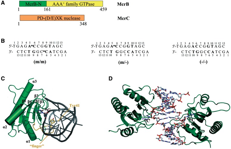
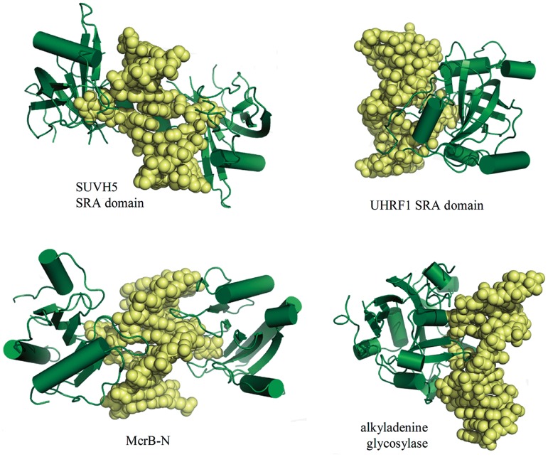
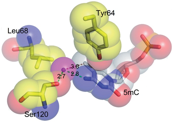
DNA cytosine methylation is a widespread epigenetic mark. Biological effects of DNA methylation are mediated by the proteins that preferentially bind to 5-methylcytosine (5mC) in different sequence contexts. Until now two different structural mechanisms have been established for 5mC recognition in eukaryotes; however, it is still unknown how discrimination of the 5mC modification is achieved in prokaryotes. Here we report the crystal structure of the N-terminal DNA-binding domain (McrB-N) of the methyl-specific endonuclease McrBC from Escherichia coli. The McrB-N protein shows a novel DNA-binding fold adapted for 5mC-recognition. In the McrB-N structure in complex with methylated DNA, the 5mC base is flipped out from the DNA duplex and positioned within a binding pocket. Base flipping elegantly explains why McrBC system restricts only T4-even phages impaired in glycosylation [Luria, S.E. and Human, M.L. (1952) A nonhereditary, host-induced variation of bacterial viruses. J. Bacteriol., 64, 557-569]: flipped out 5-hydroxymethylcytosine is accommodated in the binding pocket but there is no room for the glycosylated base. The mechanism for 5mC recognition employed by McrB-N is highly reminiscent of that for eukaryotic SRA domains, despite the differences in their protein folds.


Figures

Figure 1.
Figure 1.
(A) Schematic representation of the McrBC restriction system. McrBC is composed of two subunits: McrB harbours an N-terminal DNA-binding domain (McrB-N) and GTP-ase motifs, while McrC contains a nuclease active site (20,22,23). (B) The oligoduplexes used for crystallization. The 5′-AmC (5′-AC) sequences are shown in bold. (C) Closer view of the McrB-N monomer bound to DNA. The ‘finger’ loop penetrating into the minor groove is highlighted in orange. (D) Overall structure of the two McrB-N monomers bound to the di-methylated (m/m) oligoduplex. The DNA is bent by ∼29° and both the 5-methylcytosines are flipped out.
Figure 2.
Figure 2.
Gel mobility shift analysis for McrB-N binding to DNA. (A) McrB-N binding to the oligoduplexes used for crystallization. McrB-N at concentrations indicated above the relevant lanes was mixed with the di-methylated (m/m), hemi-methylated (m/−) or non-methylated (−/−) DNA (Figure 1B) at the final concentration of 100 nM. The samples were electrophoresed through 8% PAA gels under native conditions. (B) Dependence of the McrB-N binding on the sequence context 5′-upstream of 5mC McrB-N at concentrations indicated above the relevant lanes was mixed with the di-methylated (m/m) DNA containing either G, C or T nucleotide upstream of the 5mC. The samples were analysed as described in (A).
Figure 3.
Figure 3.
Detailed protein–DNA interactions in the McrB-N complex with di-methylated DNA. (A) Schematic representation of hydrogen bond interactions. Residues from different McrB-N monomers are shown in blue and red, water molecules are shown as red spheres. In the crystal there are two ways for the DNA duplex binding to McrB-N. Crystallographic quality parameters indicate that both binding modes are possible, and therefore both the DNA duplexes were modelled. Only one of two possible DNA orientations is shown in the picture. The protein–DNA interactions are almost identical in the McrB-N complexes with hemi-methylated and non-methylated DNA (Supplementary Figure S3). (B) Close-up stereo-view of the ‘finger’ loop penetrating into the minor DNA groove. (C) Close-up stereo-view of the McrB-N interactions with the flipped out 5mC. (D) Multiple sequence alignment of the McrB-N family proteins. NPS@ server (48) was used for analysis. Secondary structure for McrB-N is displayed above the sequence. Identical residues are indicated by red shading. Sequence names are composed of organism abbreviation and the GI number.
Figure 4.
Figure 4.
McrB-N interaction with PC DNA. (A) McrB-N binding to the oligoduplexes containing one (p/m) or two PC (p/p) bases (Table 2). The binding reactions contained 33P-labelled 30-bp oligoduplex (100 nM) and the protein at concentrations as indicated by each lane. The samples were electrophoresed through 8% PAA gels under native conditions. (B) PC steady state fluorescence measurements in solution. Left panel: corrected PC emission spectra of McrB-N–DNA complexes. Reactions contained 5 μM protein and 1 μM oligoduplex p/p or p/m (see ‘Materials and Methods’ section for the details). Right panel: diagrams illustrate the maximum fluorescence intensity (456 nm for p/p, 459 nm for McrB-N–p/p, 466 nm for p/m and 463 nm for McrB-N–p/m) values of the corrected fluorescence emission spectra presented in left panel.
Figure 5.
Figure 5.
McrB-N in comparison to other base-flipping proteins. Two monomers of SUVH5 SRA domain (3Q0C) or McrB-N are bound to the same DNA duplex, while in the case of the UHRF SRA domain (2ZKD) a single monomer is bound to the DNA. The DNA retains almost canonical B-DNA structure, except for the flipped out nucleotide, in the SRA domain complexes, while McrB-N bends DNA similarly to the human alkyladenine glycosylase (1BNK).
Figure 6.
Figure 6.
Close-up view of the water molecule in the vicinity of the methyl group in the McrB-N complex with di-methylated DNA. The hydroxyl group of 5hmC or methyl group of the N4-methylcytosine may occupy the space filled by the water molecule (shown in magenta).

Similar articles

Cited by

References

    1. Bird A. DNA methylation patterns and epigenetic memory. Genes Dev. 2002;16:6–21. - PubMed
    1. Tost J. DNA methylation: an introduction to the biology and the disease-associated changes of a promising biomarker. Mol. Biotechnol. 2010;44:71–81. - PubMed
    1. Henderson IR, Jacobsen SE. Epigenetic inheritance in plants. Nature. 2007;447:418–424. - PubMed
    1. Ho KL, McNae IW, Schmiedeberg L, Klose RJ, Bird AP, Walkinshaw MD. MeCP2 binding to DNA depends upon hydration at methyl-CpG. Mol. Cell. 2008;29:525–531. - PubMed
    1. Arita K, Ariyoshi M, Tochio H, Nakamura Y, Shirakawa M. Recognition of hemi-methylated DNA by the SRA protein UHRF1 by a base-flipping mechanism. Nature. 2008;455:818–821. - PubMed

Publication types

MeSH terms