. 2012 Jul 6;287(28):23332-45.
doi: 10.1074/jbc.M112.348839. Epub 2012 May 8.

Structural and functional insight into how the Plasmodium falciparum VAR2CSA protein mediates binding to chondroitin sulfate A in placental malaria

Affiliations

Structural and functional insight into how the Plasmodium falciparum VAR2CSA protein mediates binding to chondroitin sulfate A in placental malaria

Thomas M Clausen et al. J Biol Chem. .

Abstract

Malaria is a major global health problem. Pregnant women are susceptible to infection regardless of previously acquired immunity. Placental malaria is caused by parasites capable of sequestering in the placenta. This is mediated by VAR2CSA, a parasite antigen that interacts with chondroitin sulfate A (CSA). One vaccine strategy is to block this interaction with VAR2CSA-specific antibodies. It is a priority to define a small VAR2CSA fragment that can be used in an adhesion blocking vaccine. In this, the obvious approach is to define regions of VAR2CSA involved in receptor binding. It has been shown that full-length recombinant VAR2CSA binds specifically to CSA with nanomolar affinity, and that the CSA-binding site lies in the N-terminal part of the protein. In this study we define the minimal binding region by truncating VAR2CSA and analyzing CSA binding using biosensor technology. We show that the core CSA-binding site lies within the DBL2X domain and parts of the flanking interdomain regions. This is in contrast to the idea that single domains do not possess the structural requirements for specific CSA binding. Small-angle x-ray scattering measurements enabled modeling of VAR2CSA and showed that the CSA-binding DBL2X domain is situated in the center of the structure. Mutating classic sulfate-binding sites in VAR2CSA, along with testing dependence of ionic interactions, suggest that the CSA binding is not solely dependent on the sulfated CSA structure. Based on these novel PfEMP1 structure-function studies, we have constructed a small VAR2CSA antigen that has the capacity to induce highly adhesion-blocking antibodies.


Figures

FIGURE 1.
FIGURE 1.
Schematic representation of produced VAR2CSA fragments. ID2a-ID2b corresponds to CIDRPAM in Ref. . The figure shows the tested fragments along with KD values determined in the kinetic analysis. The kinetic analysis was performed on a Quartz Crystal Microbalance (Attana A100). The sensorgrams are shown in Fig. 3. ID1-DBL2Xb and ID1-DBL2Xa were produced based on the 3D7 genotype. The minimal binding region is ID1-DBL2Xb.
FIGURE 2.
FIGURE 2.
Screening for CSPG binding VAR2CSA fragments in a solid phase binding assay (ELISA). The protein tested is given at the top of each graph. For schematic representation of fragments, see Fig. 1. Microtiter plates were coated with 3 μg/ml of CSPG, 3 μg/ml of HSPG, and 1% BSA. A 2-fold dilution series of protein (1.56–100 nm) was added to the wells. Bound protein was detected using anti-V5 HRP-conjugated antibody. The plates were developed with OPD and absorbance was detected at 490 nm. The minimal CSPG-binding protein is ID1-DBL2Xb.
FIGURE 3.
FIGURE 3.
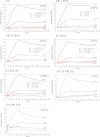
Examining binding kinetics of minimal binding fragments on the Quartz Crystal Microbalance (Attana A100). The protein tested is given at the top of each graph. For schematic representation of fragments, see Fig. 1. Sensorgrams show VAR2CSA fragments binding to immobilized CSPG. The binding is illustrated as a change in frequency over time. The CSPG was coated at 100 μg/ml. The proteins were injected at different concentrations in a 1:3 dilution series (0.25–60 μg/ml). The association and disassociation phases are shown. The black curve corresponds to the actual data collected and the red is the fitted 1:1 binding model. Values for kon, koff, and the calculated KD values are shown in each graph. It was not possible to fit a 1:1 binding model onto DBL1X-DBL2Xa or ID1-DBL2Xa. The minimal CSPG-binding protein is ID1-DBL2Xb.
FIGURE 4.
FIGURE 4.
Testing the inhibitory capacity of serum extracted in VAR2CSA fragment immunizations. The figure shows inhibition of VAR2CSA expressing FCR3-infected erythrocytes binding to CSPG using VAR2CSA-specific serum. Serum was tested at several dilutions (1:10, 1:20, and 1:50). The fraction of binding parasites (tritium labeled) was measured by liquid scintillation. Inhibition is given relative to the positive control (binding without inhibitor). The dotted line indicates the mean + 2 S.D. of binding to bovine serum albumin. We show that all CSA-binding fragments induce a strong anti-adhesive immune response in rats.
FIGURE 5.
FIGURE 5.
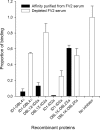
Testing the inhibitory capacity of anti-FV2 Ig, affinity purified on minimal binding fragments. The figure shows inhibition of VAR2CSA-expressing FCR3-infected erythrocytes binding to CSPG using anti-FV2 Ig affinity purified on VAR2CSA fragments. The inhibitory capacity was tested for both affinity purified samples and the depleted run-through. The samples were tested at a 1:10 dilution. The fraction of binding parasites (tritium labeled) was measured by liquid scintillation. Inhibition is given relative to the positive control (binding without inhibitor). We show that inhibitory antibodies target the minimal binding region.
FIGURE 6.
FIGURE 6.
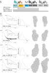
Experimental SAXS curves, pair-distance distribution functions P(r), and ab initio shape reconstructions. A, a schematic representation of VAR2CSA fragments used in the SAXS analysis together with color-coded VAR2CSA crystal structure homologues (PDB codes 1ZRO (25) and 3C64 (21)) used in the docking processes for the presentation in Fig. 7. White boxes refer to poorly homologous inter-domain linker regions of VAR2CSA (ID's) separating single DBL domains. B, left, SAXS curves for DBL1X-ID2a, ID1-DBL4ϵ, DBL1X-DBL4ϵ, and FV2 (DBL1X-DBL6ϵ). The logarithm of the scattering intensity (log(I)) is shown as a function of the momentum transfer q in units of Å−1. Gray solid lines show the fit of the shape reconstructions to the experimental data. χ2 values were 2.5, 1.3, 1.5, and 1.1, respectively. The Guinier fit are shown as the natural logarithm of the scattering intensity as a function of the squared momentum transfer. Center, P(r) functions calculated from each scattering curve are plotted in relative units as a function of the distance in nanometers. Right, the average ab initio shape reconstructions are shown with spheres representing the dummy-atoms used by DAMMIF. The orientation on the right is after a 90° right-hand rotation.
FIGURE 7.
FIGURE 7.
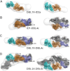
Docking of crystal structure homologues into the molecular envelopes obtained from SAXS. The molecular envelopes are depicted as a mesh. The docked crystal structure homologues are shown as domains with a surface representation. A, DBL1X-ID2a; B, ID1-DBL4ϵ; C, DBL1-DBL4ϵ; D, DBL1X-DBL6ϵ. The left panel shows a side view and the right panel shows the top view after a 90° rotation.
FIGURE 8.
FIGURE 8.
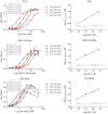
Influence of ionic strength on VAR2CSA binding to CSPG. Proteins tested were FV2, DBL1X-ID2a, and ID1-ID2a from 3D7. Left panel, ELISA-based binding assay. CSPG was coated at 3 μg/ml. A 1:2 dilution series (400 to 1.56 nm) of protein was added in several different NaCl concentrations (150, 200, 250, and 300 mm). The KD observed values were calculated for each titration series in GraphPad Prism using nonlinear regression (least squares fit with Hill slope). Fit is shown in red. Right column, log KD, observed versus log[Na+]. The graph is linear between 150 and 300 mm [NaCl]. The linear relationship is given as y = ax + b. The slope (a) corresponds to m(1-f) and b corresponds to log KD,nonionic as illustrated in Equation 2.

References

    1. WHO (2010) World Malaria Report 2010, WHO, Geneva, Switzerland
    1. Doolan D. L., Dobaño C., Baird J. K. (2009) Acquired immunity to malaria. Clin. Microbiol. Rev. 22, 13–36 - PMC - PubMed
    1. Fried M., Duffy P. E. (1996) Adherence of Plasmodium falciparum to chondroitin sulfate A in the human placenta. Science 272, 1502–1504 - PubMed
    1. Menendez C. (1995) Malaria during pregnancy. A priority area of malaria research and control. Parasitol. Today 11, 178–183 - PubMed
    1. McGregor I. A. (1987) Thoughts on malaria in pregnancy with consideration of some factors that influence remedial strategies. Parassitologia 29, 153–163 - PubMed

Publication types

MeSH terms

Substances

LinkOut - more resources