. 2012 Jun 5;109(23):9161-6.
doi: 10.1073/pnas.1202916109. Epub 2012 May 8.

Hydrogen sulfide and nitric oxide are mutually dependent in the regulation of angiogenesis and endothelium-dependent vasorelaxation

Affiliations

Hydrogen sulfide and nitric oxide are mutually dependent in the regulation of angiogenesis and endothelium-dependent vasorelaxation

Ciro Coletta et al. Proc Natl Acad Sci U S A. .

Abstract

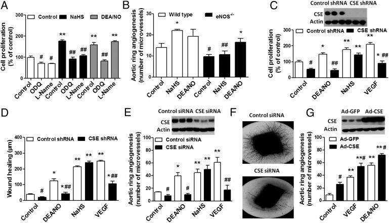
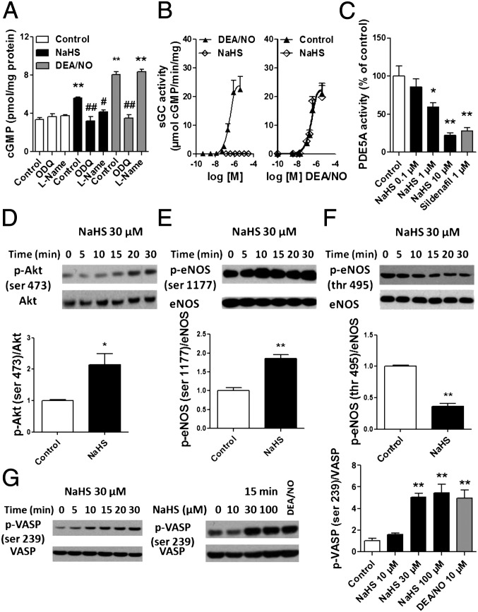
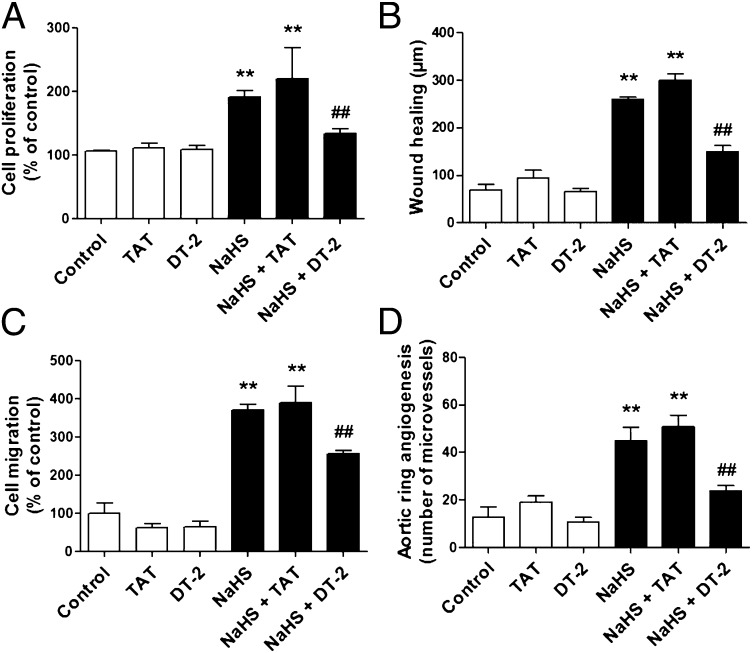
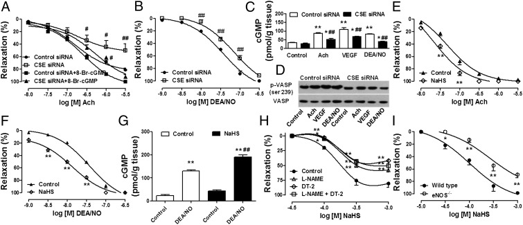
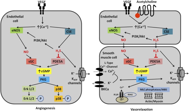
Hydrogen sulfide (H(2)S) is a unique gasotransmitter, with regulatory roles in the cardiovascular, nervous, and immune systems. Some of the vascular actions of H(2)S (stimulation of angiogenesis, relaxation of vascular smooth muscle) resemble those of nitric oxide (NO). Although it was generally assumed that H(2)S and NO exert their effects via separate pathways, the results of the current study show that H(2)S and NO are mutually required to elicit angiogenesis and vasodilatation. Exposure of endothelial cells to H(2)S increases intracellular cyclic guanosine 5'-monophosphate (cGMP) in a NO-dependent manner, and activated protein kinase G (PKG) and its downstream effector, the vasodilator-stimulated phosphoprotein (VASP). Inhibition of endothelial isoform of NO synthase (eNOS) or PKG-I abolishes the H(2)S-stimulated angiogenic response, and attenuated H(2)S-stimulated vasorelaxation, demonstrating the requirement of NO in vascular H(2)S signaling. Conversely, silencing of the H(2)S-producing enzyme cystathionine-γ-lyase abolishes NO-stimulated cGMP accumulation and angiogenesis and attenuates the acetylcholine-induced vasorelaxation, indicating a partial requirement of H(2)S in the vascular activity of NO. The actions of H(2)S and NO converge at cGMP; though H(2)S does not directly activate soluble guanylyl cyclase, it maintains a tonic inhibitory effect on PDE5, thereby delaying the degradation of cGMP. H(2)S also activates PI3K/Akt, and increases eNOS phosphorylation at its activating site S1177. The cooperative action of the two gasotransmitters on increasing and maintaining intracellular cGMP is essential for PKG activation and angiogenesis and vasorelaxation. H(2)S-induced wound healing and microvessel growth in matrigel plugs is suppressed by pharmacological inhibition or genetic ablation of eNOS. Thus, NO and H(2)S are mutually required for the physiological control of vascular function.


Conflict of interest statement

Conflict of interest statement: C.S. is a holder of patents related to the therapeutic effects of H2S.

Figures

Fig. 1.
Fig. 1.
Mutual requirement of NO and H2S for angiogenesis in vitro. (A) bEnd3 cells were seeded overnight in 12-well plates, pretreated with ODQ (10 μM, 2 h) or l-NAME (4 mM, 40 min), and subsequently incubated with NaHS (30 μM) or DEA/NO (10 μM) for an additional 48 h. Cells were trypsinized and counted using a Neubauer hemocytometer. **P < 0.01 vs. control; ##P < 0.01 vs. NaHS. (B) Aortic rings were harvested from wild-type or eNOS−/− mice, cultured for 7 d in collagen gel in Opti-MEM medium containing 1% FBS, in the presence or absence of NaHS (30 μM) or DEA/NO (10 μM). *P < 0.05 vs. corresponding control; #P < 0.05 and ##P < 0.01 vs. corresponding wild type. (C) The lentiviral shRNA vector targeting CSE was transfected into bEnd3 cells. The shRNA vector effectively inhibited the expression of CSE gene in bEnd3 cell line at the protein level, as shown by Western blot analysis (C Upper). Following CSE silencing, cells were seeded in 12-well cell culture plates, and cell proliferation (C) and wound healing (D) were then evaluated in the presence of vehicle, DEA/NO (10 μM), NaHS (30 μM), or VEGF (20 ng/mL). *P < 0.05 and **P < 0.01 vs. control; #P < 0.05 and ##P < 0.01 vs. corresponding control shRNA. (E) Rat aortic rings were silenced with CSE siRNA for 48 h. Following gene silencing, aortic explants were placed individually on the bottom of 24-well plates, and collagen gel was gently applied. Rings were cultured for 7 d in the presence of vehicle, DEA/NO (10 μM), NaHS (30 μM), or VEGF (20 ng/mL). Western blots confirm efficient CSE silencing in aortic rings. CSE silencing markedly reduced both VEGF- and DEA/NO-induced vessel sprouting. *P < 0.05 and **P < 0.01 vs. control; #P < 0.05 and ##P < 0.01 vs. corresponding control siRNA. (F) Representative images of the 7-d collagen gel cultures of aortic rings exposed to VEGF following CSE gene silencing. (G) Aortic rings were exposed to adenovirus expressing GFP or CSE before embedding in collagen gel. Overexpression of CSE enhanced VEGF- or DEA/NO-induced neovessel growth. **P < 0.01 vs. control; #P < 0.05 vs. corresponding Ad-GFP. (Upper) Representative Western blot for CSE protein in rings exposed to adenovirus expressing GFP or CSE.
Fig. 2.
Fig. 2.
H2S and NO signaling converge on the cGMP/PKG pathway. (A) bEnd3 cells were pretreated with l-NAME (4 mM, 40 min) or ODQ (10 μM, 2 h). Following pretreatments, cells were washed twice with Hanks’ balanced salt solution and then exposed for 3 min to NaHS at the indicated concentrations. cGMP was extracted in 0.1 M HCl and measured by enzyme immunoassay. **P < 0.01 vs. control; #P < 0.05 and ##P < 0.01 vs. corresponding control. (B) sGC enzymatic activity was assayed using [α-32P]GTP to [32P]cGMP conversion assay in response to the indicated concentrations of NaHS or DEA/NO. To test whether H2S affects NO-induced sGC activation, the effect of increasing concentrations of DEA/NO on sGC activity was assessed in the presence or absence of 10 μM NaHS. (C) PDE5A activity was measured in the presence of the indicated concentrations of NaHS or 1 μM sildenafil, as a positive control. *P < 0.05 or **P < 0.01 vs. control. bEnd3 cells were exposed to NaHS at the indicated concentrations and time. Cell lysates were analyzed by SDS/PAGE. PVDF membranes were blotted by using rabbit polyclonal antibodies against phosphorylated (Ser473) or total Akt (D); phosphorylated (Ser1177) or total eNOS (E); phosphorylated (Thr495) or total eNOS (F); and phosphorylated (Ser239) or total VASP (G). Densitometric analysis was performed on three blots from three different experiments using image analysis software. *P < 0.05 and **P < 0.01 NaHS (30 min) vs. control.
Fig. 3.
Fig. 3.
PKG-I inhibition prevents H2S-induced in vitro angiogenesis. (A) bEnd3 cells were pretreated with vehicle, DT-2 peptide (1 μM, 20 min), or the control peptide (TAT) (1 μM, 20 min) and then stimulated with NaHS (30 μM) to induce cell proliferation. The effect of DT-2 on NaHS-induced response was also estimated in wound healing (B) and cell migration (C). **P < 0.01 vs. control; ##P < 0.01 vs. NaHS + TAT. (D) Aortic ring explants were embedded in collagen gel, treated with the control peptide (TAT) or DT-2 (1 μM), and cultured for 7 d in the presence or absence of NaHS (30 μM, applied every 8 h). P < 0.01 vs. control; ##P < 0.01 vs. NaHS + TAT.
Fig. 4.
Fig. 4.
In vivo relevance for the interdependence of H2S and NO on angiogenesis. (A) The effect of H2S on angiogenesis in vivo was assessed by Matrigel plug assay in wild-type or eNOS−/− mice. Neovascularization in the matrigel plugs was quantified by measuring hemoglobin content using Drabkin’s reagent. NaHS treatments (50 μmol/kg per day) significantly promoted neovascularization in the Matrigel plugs in wild-type mice. The effect of NaHS treatment was abolished in mice lacking eNOS. **P < 0.01 vs. control; ##P < 0.01 vs. corresponding wild type. (B) Images of the Matrigel plugs immediately after collection from wild-type and eNOS−/− mice. (C) Following anesthesia and analgesia, rats were placed in a mold and subjected to burn injury by submerging the back in scalding (96–99 °C) water for 10 s. Rats were randomly divided into four groups and treated daily for 28 d either with vehicle or NaHS (300 μg/kg per day s.c.). Rats from l-NAME groups received daily s.c. injections of the eNOS inhibitor (20 mg/kg). Subcutaneous injections of NaHS were performed twice daily at four equally spaced sites in the transition zone between burn site and healthy tissue. Burn wound area was determined on day 28 by planimetry. *P < 0.05 vs. control; ##P < 0.01 vs. corresponding control. (D) Representative images of the skin wounds at 28 d.
Fig. 5.
Fig. 5.
Requirement of simultaneous production of H2S and NO in vasorelaxation. Cumulative-concentration response curves to acetylcholine (A) or DEA/NO (B) were performed in aortic rings incubated in isolated organ baths following siRNA-mediated silencing of CSE. ##P < 0.01 vs. control siRNA. To test the effect of cGMP elevation on acetylcholine induced vasorelaxation, cell-permeable cGMP (8-Br-cGMP) was applied at 10 μM concomitantly to cumulative acetylcholine administrations. (C) Aortic rings subjected to CSE gene silencing were stimulated with acetylcholine (1 μM), DEA/NO (0.1 μM), or VEGF (50 ng/mL) for 15 min. At the end of the incubation time, cGMP was extracted and measured by enzyme immunoassay. **P < 0.01 vs. control; ##P < 0.01 vs. corresponding control siRNA. (D) Following CSE gene targeting (siRNA), aortic rings were incubated in Krebs–Henseleit buffer at 37 °C and stimulated with acetylcholine (1 μM, 15 min), DEA/NO (0.1 μM, 15 min), or VEGF (50 ng/mL, 15 min). Tissue lysates were analyzed by SDS/PAGE. PVDF membranes were blotted by using rabbit polyclonal antibodies against phosphorylated (ser 239) or total VASP. (E) Aortic rings were incubated with NaHS 30 μM on PE-induced stable tone. Concentration response curves to acetylcholine (E) or DEA/NO (F) were performed. In these settings, NaHS enhances the vasorelaxant properties of endothelium-derived or exogenously applied NO. **P < 0.01 vs. control. Similarly, 15-min exposure of aortic rings to a low concentration of NaHS (30 μM) enhanced DEA/NO-induced elevation of intracellular cGMP as assessed by enzyme immunoassay (G). (H) Aortic rings were mounted in organ baths, pretreated with vehicle, l-NAME (100 μM), DT-2 (1 μM), or l-NAME + DT-2 on basal tone for 30 min. Rings were then challenged with PE (1 μM), and concentration response curves to NaHS were performed on stable tone. *P < 0.05 and *P < 0.01 vs. control. (I) Aortic tissues were dissected from wild-type and eNOS−/− mice and placed in isolated organ baths under a resting tension of 1.5 g. Following a stabilization period, aortic rings were challenged with PE (1 μM), and cumulative concentration response curves to NaHS were performed. *P < 0.05 and **P < 0.01 vs. wild type.
Fig. 6.
Fig. 6.
Proposed pathways of H2S and NO interaction. Cooperation between NO and H2S in angiogenesis (Left) or endothelium-dependent vasorelaxation (Right). Binding of VEGF or acetylcholine to its receptor on the endothelial cell mobilizes intracellular calcium and activates eNOS as well as CSE (two calcium-dependent enzymes), resulting in a simultaneous elevation of intracellular NO and H2S levels in endothelial cells (in the context of angiogenesis) or in the smooth muscle cell (in the context of vasorelaxation). NO stimulates guanylyl cyclase, whereas endogenously produced H2S maintains a tonic inhibitory effect on PDE5, thereby delaying the degradation of cGMP and permitting physiological cGMP signaling. These two simultaneous actions ensure that cGMP has a sufficiently long half-life to activate PKG to stimulate PKG-dependent downstream signaling such as ERK1/2 and p38 in the case of angiogenesis and myosin light chain (MLC) phosphatase (via its myosin-binding subunit, MBS), large-conductance calcium- and voltage-activated potassium channels (BKCa), and IP3-R-–associated cG-kinase substrate (IRAG) in the case of smooth muscle relaxation. NO and H2S are also known to activate KATP channels, which are also involved in angiogenesis and endothelium-dependent relaxation.

Comment in

  • Shared signaling pathways among gasotransmitters.
    Wang R. Wang R. Proc Natl Acad Sci U S A. 2012 Jun 5;109(23):8801-2. doi: 10.1073/pnas.1206646109. Epub 2012 May 21. Proc Natl Acad Sci U S A. 2012. PMID: 22615409 Free PMC article. No abstract available.

References

    1. Mustafa AK, Gadalla MM, Snyder SH. Signaling by gasotransmitters. Sci Signal. 2009;2:re2. - PMC - PubMed
    1. Szabo C. Gaseotransmitters: New frontiers for translational science. Sci Transl Med. 2010;2:59ps54. - PMC - PubMed
    1. Ignarro LJ. Nitric oxide: A unique endogenous signaling molecule in vascular biology. Biosci Rep. 1999;19:51–71. - PubMed
    1. Bryan NS, Bian K, Murad F. Discovery of the nitric oxide signaling pathway and targets for drug development. Front Biosci. 2009;14:1–18. - PubMed
    1. Wang R. The gasotransmitter role of hydrogen sulfide. Antioxid Redox Signal. 2003;5:493–501. - PubMed

Publication types

MeSH terms

LinkOut - more resources