. 2012;8(5):e1002667.
doi: 10.1371/journal.ppat.1002667. Epub 2012 May 3.

The lipopolysaccharide from Capnocytophaga canimorsus reveals an unexpected role of the core-oligosaccharide in MD-2 binding

Affiliations

The lipopolysaccharide from Capnocytophaga canimorsus reveals an unexpected role of the core-oligosaccharide in MD-2 binding

Simon Ittig et al. PLoS Pathog. 2012.

Abstract

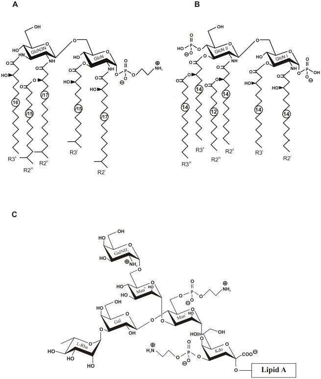
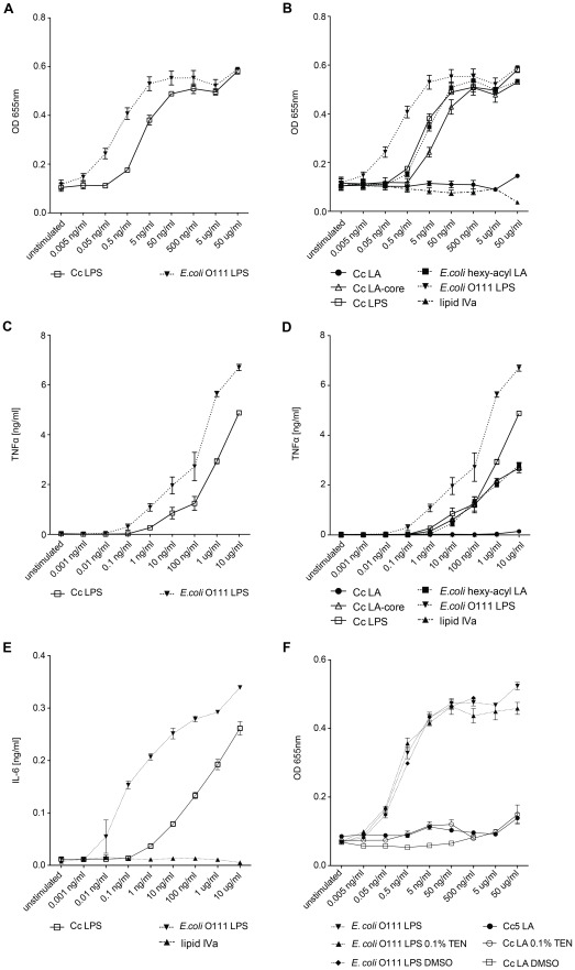
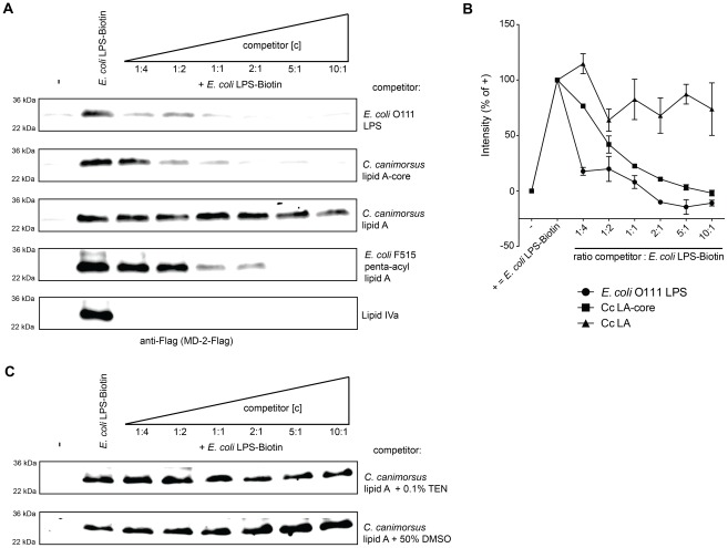
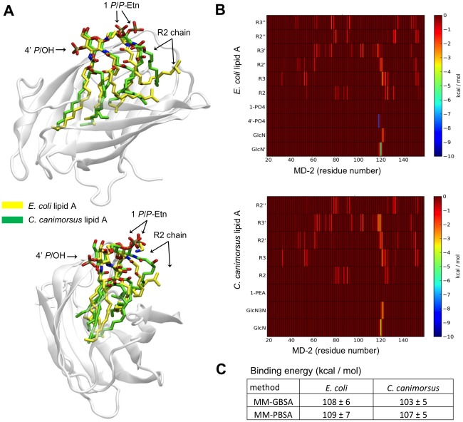
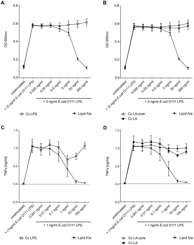
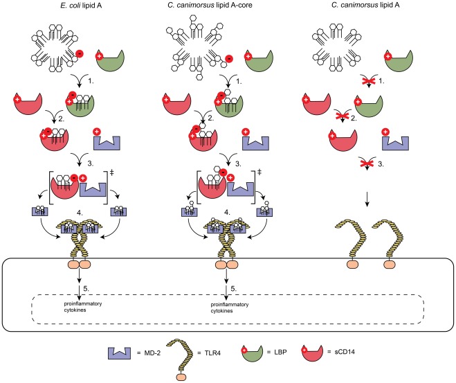
Capnocytophaga canimorsus is a usual member of dog's mouths flora that causes rare but dramatic human infections after dog bites. We determined the structure of C. canimorsus lipid A. The main features are that it is penta-acylated and composed of a "hybrid backbone" lacking the 4' phosphate and having a 1 phosphoethanolamine (P-Etn) at 2-amino-2-deoxy-d-glucose (GlcN). C. canimorsus LPS was 100 fold less endotoxic than Escherichia coli LPS. Surprisingly, C. canimorsus lipid A was 20,000 fold less endotoxic than the C. canimorsus lipid A-core. This represents the first example in which the core-oligosaccharide dramatically increases endotoxicity of a low endotoxic lipid A. The binding to human myeloid differentiation factor 2 (MD-2) was dramatically increased upon presence of the LPS core on the lipid A, explaining the difference in endotoxicity. Interaction of MD-2, cluster of differentiation antigen 14 (CD14) or LPS-binding protein (LBP) with the negative charge in the 3-deoxy-D-manno-oct-2-ulosonic acid (Kdo) of the core might be needed to form the MD-2 - lipid A complex in case the 4' phosphate is not present.


Conflict of interest statement

The authors have declared that no competing interests exist.

Figures

Figure 1
Figure 1. NMR analysis of the lipid A from C. canimorsus wild type.
1H,13C-HSQC spectrum (700 MHz) of lipid A in chloroform-methanol-water (20∶10∶1, v/v/v) at 27°C. The corresponding parts of the 13C and 1H NMR spectra are displayed along the F1 and F2 axes, respectively. Numerals refer to atoms in sugar and acyl chain residues denoted by letters as shown in Supplementary Table 1 and Fig. S2. Signals from an unidentified contaminating lipid are indicated by X.
Figure 2
Figure 2. Structures of C. canimorsus lipid A, E. coli lipid A and core-oligosaccharide of C. canimorsus attached to the lipid A.
(A) C. canimorsus lipid A consists of a β-(1′→6)-linked GlcN3N′-GlcN disaccharide, to which 3-hydroxy-15-methylhexadecanoic acid, 3-hydroxy-13-methyltetradecanoic acid, 3-O-(13-methyltetradecanoyl)-15-methylhexadecanoic acid, and 3-hydroxyhexadecanoic acid are attached at positions 2, 3, 2′, and 3′, respectively. The disscharide carries a positively charged ethanolamine at the 1 phosphate and lacks a 4′ phosphate. (B) Structure of E. coli hexa-acylated lipid A. (C) C. canimorsus LPS core features only one Kdo, to which a phosphoethanolamine (P-Etn) is attached. The only net negative charge present is from the carboxy group of the Kdo. The inner core continues with Man to which another a P-Etn is attached. The outer core consists of Gal and l-Rhamnose (l-Rha), to which the O-antigen is attached (U. Zähringer, unpublished results).
Figure 3
Figure 3. Biosynthesis of C. canimorsus lipid A-Kdo.
(A) Alphabetic list of enzymes required and the corresponding gene codes in the C. canimorsus 5 genome are listed. (B) Proposed enzymatic modification on lipid A by LpxF, LpxE and EptA. (C) Single steps in the biosynthesis of C. canimorsus lipid A-Kdo (adapted from KEGG map00540).
Figure 4
Figure 4. Endotoxic activity of C. canimorsus (Cc) LPS, lipid A (LA) or lipid A-core (LA-core) and contribution of the LPS core to endotoxicity.
(A–B) Dose-response curve of purified lipid A, LA-core or LPS. Samples were assayed for TLR4 dependent NFκB activation with HEKBlue human TLR4 cells. (C–D) Purified lipid A, LA-core or LPS samples were assayed for induction of TNFα release by human THP-1 macrophages. (E) Purified LPS samples or lipid IVA were assayed for induction of IL-6 release by canine DH82 macrophages. (F) Dose-response curve of NFκB activation by lipid A or LPS. Purified lipid A or LPS samples were assayed for TLR4-dependent NFκB activation with HEKBlue human TLR4 cells. The C. canimorsus lipid A stock solution was pretreated with either 0.1% TEN or 50% DMSO and sonication to increase its solubility in water/buffer. Identical concentrations of DMSO or TEN were added to E. coli O111 LPS as a control. Data were combined from n = 3 independent experiments, error bars indicated are standard error of the mean.
Figure 5
Figure 5. Binding to human MD-2 of C. canimorsus lipid A depends on the core-oligosaccharide.
Soluble human MD-2 from cell culture supernatant was combined with the indicated mixture of E. coli LPS-Biotin and a competitor (either C. canimorsus lipid A, lipid A-core, E. coli O111 LPS, penta-acyl E. coli lipid A or lipid IVA). Biotinylated E. coli LPS-MD-2 complexes were purified and analyzed by non-reducing, denaturing Western blotting for presence of MD-2. (A) Untreated human MD-2 did not bind to the Strep-column (lane 1), addition of E. coli LPS-biotin lead to co-purification of human MD-2 (lane 2). Results shown are representative of three independent determinations. (B) Quantification of Western-blots as shown in A. Values are shown as percentage of the corresponding positive control. Data were combined from n = 3 independent experiments, error bars indicated are standard error of the mean. (C) as in (A) but the C. canimorsus lipid A stock solution was pretreated with either 0.1% TEN or 50% DMSO and sonication in both cases to increase its solubility in water/buffer.
Figure 6
Figure 6. Modeled binding of C. canimorsus lipid A to human MD-2.
(A) Front and side view of the equilibrated complexes between MD-2 (gray) and C. canimorsus (yellow) and E. coli (green) lipid A. (B) Pairwise decomposition of the global total (Van der Waals+electrostatic+solvation) binding free energy calculated at MM-GBSA level. (C) Binding energy between MD-2 and the two lipid A molecules calculated using the MM-GBSA and MM-PBSA methods on 300 snapshots extracted from two 10 ns long equilibrated NPT molecular dynamics simulations.
Figure 7
Figure 7. Antagonistic activity of C. canimorsus (Cc) LPS, lipid A (LA) or LA-core on the action of E. coli O111 LPS.
(A–B) HEKBlue human TLR4 cells were preincubated for 3 h with purified lipid A, LA-core or LPS samples at the concentration indicated. Then the cells were stimulated with 5 ng/ml E. coli O111 LPS for further 20–24 h and TLR4 dependent NFκB activation was measured. (C–D) Human THP-1 macrophages were preincubated for 3 h with purified lipid A, LA-core or LPS samples at the concentration indicated. Then the cells were stimulated with 1 ng/ml E. coli O111 LPS for further 20 h and TNFα release was measured. Data were combined from n = 3 independent experiments, error bars indicated are standard error of the mean.
Figure 8
Figure 8. Proposed model for the implication of the LPS core or the 4′ phosphate in enabling the binding to MD-2.
Ionic interactions or hydrogen bonds involving the 4′ phosphate or the Kdo carboxy group in LPS lacking a 4′ phosphate enable the binding of lipid A to either LBP (1.), soluble CD14 (sCD14) (2.) or via an intermediate state to MD-2 (3.). Dependent on the type of lipid A bound to MD-2 this leads to TLR4 multimerization (4.), a downstream signaling cascade and finally release of proinflammatory cytokines (5.).

References

    1. Blanche P, Bloch E, Sicard D. Capnocytophaga canimorsus in the oral flora of dogs and cats. J Infect. 1998;36:134. - PubMed
    1. Bobo RA, Newton EJ. A previously undescribed gram-negative bacillus causing septicemia and meningitis. Am J Clin Pathol. 1976;65:564–569. - PubMed
    1. Janda JM, Graves MH, Lindquist D, Probert WS. Diagnosing Capnocytophaga canimorsus infections. Emerg Infect Dis. 2006;12:340–342. - PMC - PubMed
    1. Brenner DJ, Hollis DG, Fanning GR, Weaver RE. Capnocytophaga canimorsus sp. nov. (formerly CDC group DF-2), a cause of septicemia following dog bite, and C. cynodegmi sp. nov., a cause of localized wound infection following dog bite. J Clin Microbiol. 1989;27:231–235. - PMC - PubMed
    1. Manfredi P, Pagni M, Cornelis GR. Complete Genome Sequence of the Dog Commensal and Human Pathogen Capnocytophaga canimorsus Strain 5. J Bacteriol. 2011;193:5558–5559. - PMC - PubMed

Publication types

MeSH terms