. 2012;8(4):e1002646.
doi: 10.1371/journal.pgen.1002646. Epub 2012 Apr 26.

Sequence-specific targeting of dosage compensation in Drosophila favors an active chromatin context

Affiliations

Sequence-specific targeting of dosage compensation in Drosophila favors an active chromatin context

Artyom A Alekseyenko et al. PLoS Genet. 2012.

Erratum in

Abstract

The Drosophila MSL complex mediates dosage compensation by increasing transcription of the single X chromosome in males approximately two-fold. This is accomplished through recognition of the X chromosome and subsequent acetylation of histone H4K16 on X-linked genes. Initial binding to the X is thought to occur at "entry sites" that contain a consensus sequence motif ("MSL recognition element" or MRE). However, this motif is only ∼2 fold enriched on X, and only a fraction of the motifs on X are initially targeted. Here we ask whether chromatin context could distinguish between utilized and non-utilized copies of the motif, by comparing their relative enrichment for histone modifications and chromosomal proteins mapped in the modENCODE project. Through a comparative analysis of the chromatin features in male S2 cells (which contain MSL complex) and female Kc cells (which lack the complex), we find that the presence of active chromatin modifications, together with an elevated local GC content in the surrounding sequences, has strong predictive value for functional MSL entry sites, independent of MSL binding. We tested these sites for function in Kc cells by RNAi knockdown of Sxl, resulting in induction of MSL complex. We show that ectopic MSL expression in Kc cells leads to H4K16 acetylation around these sites and a relative increase in X chromosome transcription. Collectively, our results support a model in which a pre-existing active chromatin environment, coincident with H3K36me3, contributes to MSL entry site selection. The consequences of MSL targeting of the male X chromosome include increase in nucleosome lability, enrichment for H4K16 acetylation and JIL-1 kinase, and depletion of linker histone H1 on active X-linked genes. Our analysis can serve as a model for identifying chromatin and local sequence features that may contribute to selection of functional protein binding sites in the genome.


Conflict of interest statement

The authors have declared that no competing interests exist.

Figures

Figure 1
Figure 1. Active chromatin context and elevated GC content are associated with functional MSL recognition elements (MREs) on the X chromosome, independent of MSL binding.
(A) The sequence motif for the MRE. (B–D) The average enrichment of various chromatin modifications or chromatin-binding proteins around (+/−5 kb) the functional and non-functional MREs is visualized using a heat map in each of three Drosophila cell lines: S2 in panel B; BG3 in panel C; and Kc in panel D. (E) A heat map showing the average GC content around each class of MREs. The data were obtained from genome-wide ChIP-chip profiles generated as part of the modENCODE project. Many active marks are broadly enriched around functional MREs in both male and female cell lines, suggesting that active chromatin is strongly associated with functional MREs independent of MSL binding. See also Figures S1 and S2.
Figure 2
Figure 2. Ectopic upregulation of MSL2 by Sxl RNAi treatment induces dosage compensation of X-linked genes in female Kc cells by preferentially targeting MREs in an active chromatin context.
(A) Distribution of gene expression ratios after Sxl knockdown in X chromosome and autosomes compared to control. The y-axis (density) represents the scaled proportion of the number of genes for a given log2 expression ratio (x-axis). Repression of Sxl leads to ectopic expression of MSL2, which results in dosage compensation of the X chromosome. (B) Distribution of gene expression ratios after Sxl and MSL2 double knockdown for X chromosome and autosomes compared to control. In the absence of functional MSL2, no dosage compensation is observed. (C) A heat map showing the average enrichment of H4K16 acetylation (H4K16ac) around (+/−5 kb) the functional and non-functional MREs in control (GFP) and after two independent Sxl RNAi knockdowns. The gene expression data in panels A and B are based on the same cells as replicate 1 in the heat map.
Figure 3
Figure 3. Chromatin context is predictive of functional MREs.
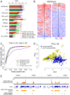
(A) A bar plot showing the performance of individual chromatin features for distinguishing functional and non-functional MREs using various training and testing schemes. The best features are H3K36me3 and JIL1. H4K16ac shows strong association with the presence of functional MREs in both male cell lines but much weaker association in female Kc cells, supporting the known role of H4K16ac as a key consequence of MSL binding. (B) A heat map showing the distribution of the H3K36me3 mark around individual functional and non-functional MREs on the X chromosome in female Kc cells, ordered by the level of enrichment at the MREs. (C) Sensitivity (y-axis) and 1 - specificity (x-axis) of prediction using a support vector machine (SVM) are shown using a receiver operator characteristic (ROC) curve. This indicates that chromatin features in female Kc cells can be used to predict the identity of functional MREs in male S2 cells with high sensitivity (high true positive rate) and specificity (low false positive rate). (D) A principal component projection of the functional and non-functional MREs based on the best chromatin features in S2 cells, showing that the two MREs groups can be well-separated. See also Figure S3. (E) A genome browser view that shows the overlap of SVM-predicted functional MREs and MSL binding sites identified by an independent MSL3 ChIP-seq analysis.
Figure 4
Figure 4. Increased lability of nucleosomes at chromatin entry sites correlates with MSL binding.
Average enrichment profiles of chromatin properties around MREs are shown for S2 and Kc cells. Nucleosome density and successive salt extracted fractions of MNase-treated chromatin from S2 and Kc cells were described previously . Profiled chromatin properties comprise nucleosome density (black) and successive salt extracted fractions of MNase-treated chromatin: low salt (pink, 80 mM NaCl), high salt (blue, 600 mM NaCl), and pellet (green, salt insoluble). The dashed red lines on each plot indicate the MRE centers. In male S2 cells, nucleosomes are more labile at the MREs on the X chromosome in general, and nucleosome density is especially low at functional MREs. The relative lability of nucleosomes on the X chromosome is not seen in female Kc cells, and the extent of nucleosome depletion at functional MREs is less pronounced. See also Figure S4.
Figure 5
Figure 5. The bodies of active X-linked genes are enriched for JIL-1 kinase and depleted for histone H1 in S2 but not Kc cell lines.
Each panel shows the average scaled ChIP enrichment profile (meta-gene profile) of active and inactive genes located on X chromosome and autosomes. (A) JIL-1 in S2 cells, (B) JIL-1 in Kc cells, (C) H1 in S2 cells, and (D) H1 in Kc cells. Notably, JIL-1 is enriched along the gene bodies of X-linked genes in males, while histone H1 is depleted. See also Figure S5.
Figure 6
Figure 6. MSL1 and H4K16 acetylation are found on virtually all active X linked genes in male S2 cells.
The plot shows the positions of exons, and the regions of enrichment for MSL1, MOF acetyltransferase, H4K16ac, and H3K36me3 along the bodies of active X linked genes. Each row represents an active gene scaled to the same size. The genes were clustered based on the chromatin features. These profiles show that CES (red dot on the H3K36me3 map) are located closer to the 3′ end in general and are embedded within domains enriched for H3K36me3 as well as MSL1, MOF, and H4K16ac.
Figure 7
Figure 7. Model for binding site selection by a chromatin associated factor.
Our results support roles for local chromatin environment and flanking GC content in discrimination of true target sites of the MSL dosage compensation complex. The model depicts the GC content and active chromatin marks surrounding MREs in female Kc cells that predict binding by MSL complex in male S2 or BG3 cells (or after MSL induction in female Kc cells). MREs that do not pre-exist in a favorable environment are not bound by MSL complex and thus are non-functional. Definition of the favorable chromatin features that pre-exist factor binding may be a general tool, in addition to DNA motif analysis, for prediction of functional binding sites.

References

    1. Amrein H, Axel R. Genes expressed in neurons of adult male Drosophila. Cell. 1997;88:459–469. - PubMed
    1. Lucchesi JC. The structure-function link of compensated chromatin in Drosophila. Cur Opin Genet Dev. 2009;19:550–556. - PMC - PubMed
    1. Straub T, Becker PB. Transcription modulation chromosome-wide: universal features and principles of dosage compensation in worms and flies. Curr Opin Genet Dev. 2011;21:147–153. - PubMed
    1. Georgiev P, Chlamydas S, Akhtar A. Drosophila dosage compensation: Males are from Mars, females are from Venus. Fly. 2011;5:147–154. - PMC - PubMed
    1. Gelbart ME, Kuroda MI. Drosophila dosage compensation: A complex voyage to the X chromosome. Development. 2009;136:1399–1410. - PMC - PubMed

Publication types

MeSH terms