. 2011 Dec;2(1):1-20.
doi: 10.1159/000332228. Epub 2011 Oct 12.

Osteogenesis Imperfecta: A Review with Clinical Examples

Affiliations

Osteogenesis Imperfecta: A Review with Clinical Examples

F S van Dijk et al. Mol Syndromol. 2011 Dec.

Abstract

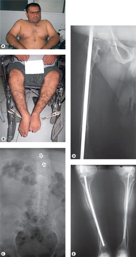
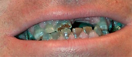
Osteogenesis imperfecta (OI) is characterized by susceptibility to bone fractures, with a severity ranging from subtle increase in fracture frequency to prenatal fractures. The first scientific description of OI dates from 1788. Since then, important milestones in OI research and treatment have, among others, been the classification of OI into 4 types (the 'Sillence classification'), the discovery of defects in collagen type I biosynthesis as a cause of most cases of OI and the use of bisphosphonate therapy. Furthermore, in the past 5 years, it has become clear that OI comprises a group of heterogeneous disorders, with an estimated 90% of cases due to a causative variant in the COL1A1 or COL1A2 genes and with the remaining 10% due to causative recessive variants in the 8 genes known so far, or in other currently unknown genes. This review aims to highlight the current knowledge around the history, epidemiology, pathogenesis, clinical/radiological features, management, and future prospects of OI. The text will be illustrated with clinical descriptions, including radiographs and, where possible, photographs of patients with OI.


Figures

Fig. 1
Fig. 1
Collagen type I biosynthesis. rER = Rough endoplasmic reticulum; Golgi = Golgi apparatus; PM = plasma membrane; ECM = extracellular matrix.
Fig. 2
Fig. 2
A Compact and spongy bone. B Endochondral ossification. Endochondral ossification occurs when mesenchymal cells differentiate into chondroblasts that produce a cartilage matrix. This cartilage acquires the shape of the bone that will be formed. A periosteal collar of bone forms around the diaphysis. Osteoblasts in this region are engaged in periosteal bone formation, which is responsible for the growth in thickness of long bones. In the centre of the diaphysis chondroblasts hypertrophy, the cartilage matrix becomes calcified, blood vessels and connective tissue cells evade the calcified cartilage, creating a marrow cavity (primary ossification center). Trabeculae of calcified cartilage (primary and secondary spongiosa) remain at the 2 ends of the cavity on which endochondral bone forms (secondary ossification centers). CHistologic abnormalities in OI compared to a control. Panels a, c and e show histology of a normal control femur at 18 weeks of gestational age (GA). Panels b, d and f are from an 18-week GA type II OI case. The transition zone of cartilage to primary spongiosa is sharp in both cases (a, b). Minor disruption can occur due to fractures and scar formation in the primary spongiosa (not shown). Panel d shows extensive metaplastic cartilage formation at the sight of a fracture. The bony trabeculae are hypercellular and the marrow is fibrotic. Panel e shows normocellular trabeculae and hematopoietic marrow between these trabeculae. In the severe OI cases, the trabeculae are thin, irregular and hypercellular in comparison with normal trabeculae (f). The marrow in between is fibrotic with hardly any hematopoiesis (d, f).
Fig. 3
Fig. 3
A Clinical pictures of patients with OI type I. Blue sclerae in a mother and daughter with OI type I. Clinical synopsis: An Iranian mother and daughter were referred because of suspected OI. The mother, 28 years old, has had 3 fractures in the wrist, ankle and femur occurring between 2–3 years of age. She developed hearing loss at the age of 13–14. She has strikingly blue sclerae, her height is 160 cm (−1.5 SD) and she has no evidence of dentinogenesis imperfecta (DI). The daughter, 8 years old, has had no fractures, a normal height of 125 cm (−1 SD) and no DI or hearing loss. Only deep blue sclerae can be observed. Coincidentally, she appears to have decreased vision due to deterioration of cone cells. Radiographs and bone densitometry of both mother and daughter were normal. There were no family members affected with OI. Molecular analyses of the COL1A1/2 genes revealed a heterozygous variant in the COL1A1 gene creating a premature stop codon (c.1081C>T; p.Arg361X). B Radiographs of a patient with OI type I. a Lateral radiograph showing osteopenia and mid thoracic vertebral compression fracture (open arrow). Note a general decrease in height, compared to adjacent vertebral bodies, and some wedging. b Oblique mid-diaphyseal fibula fracture (open arrow) and a communitive diaphyseal tibia fracture (arrow). Clinical synopsis: The patient is a 10-year-old girl with OI type I due to a deletion of the whole COL1A1 allele, who had 8 fractures so far. A fracture of the femur was noted shortly after birth. Her length is 128.5 cm (−2.5 SD), head circumference is 53.5 cm (0 SD) and bone density of the hips and lumbar vertebral column is between −4 and −7 SD, as measured by dual X-ray absorptiometry. She has wormian bones, a scoliosis and grey-blue sclerae typical for OI. She has no DI [van Dijk et al., 2010a].
Fig. 4
Fig. 4
A OI type II-A in a preterm infant. Skeletal overviews without (a) and with (b) silver nitrate impregnation show generalized osteopenia with diminished ossification of the calvarian bones and gross skeletal deformation. No vertebral anomalies are seen. The ribs are broad with continuous fractures (continuous beading). The long bones show multiple fractures and are shortened, deformed and broadened. Clinical synopsis: It concerned the second pregnancy of a nonconsanguineous Caucasian couple. At a gestational age of 21 weeks, ultrasonographic abnormalities were seen. All long bones were severely shortened (<p5). Bowing of the humeri and the femora was observed. The skull appeared somewhat doligocephalic with a remarkable clear imaging of the cerebrum and could be deformed by pressure of the ultrasound transducer. The parents decided to terminate the pregnancy, and a child was born at a gestational age of 22+3 weeks with a birth weight of 160 g. A skeletal overview revealed an almost total absence of skull mineralization and multiple fractures of the ribs and the long bones consistent with a diagnosis of OI type II-A. Bone histology showed hypercellular irregular trabeculae with multiple fractures, fibrotic marrow and metaplastic cartilage formation. On collagen electrophoresis, post-translational overmodification was observed. A causative variant in the COL1A1 gene was found (c.2300G>A; p.Gly767Asp). B Perinatal OI type II-A. a Skeletal overview of a 35+4-week-old fetus with OI type II-A shows multiple fractures of both the long bones as well as the ribs. Note the under mineralization of the skull and its deformity on the left side. b Detail of the lower extremities shows multiple consolidated fractures resulting in deformed growth of the femurs, tibiae and fibulae. c Lateral radiograph of the lumbar spine shows mild platyspondyly (arrow). Clinical synopsis: It concerned the first pregnancy of a nonconsanguineous Caucasian couple. At a pregnancy duration of 35+4 weeks, a boy was delivered by caesarean section. Apgar scores were 4/4/4 after 1, 5 and 10 min, respectively. Birth weight was 1,100 g. Blue sclerae with proptosis were visible. The thorax was small and bell-shaped. Fractures of the ribs were felt. Gasping breath with insufficient thorax excursions was apparent. Auscultation of the heart revealed bradycardia with no other anomalies. The skin was thin and fragile with a large skin defect temporo-occipital. The extremities were short and deformed. The child died 30 min after birth due to severe respiratory and circulatory insufficiency. A skeletal overview was consistent with a diagnosis of OI. Histology showed decreased osteoid formation at the level of the primary spongiosis in combination with decreased numbers of osteoblasts and osteoclasts. A causative variant in the COL1A1 gene c.1804G>A; p.Gly602Arg was found.
Fig. 4
Fig. 4
A OI type II-A in a preterm infant. Skeletal overviews without (a) and with (b) silver nitrate impregnation show generalized osteopenia with diminished ossification of the calvarian bones and gross skeletal deformation. No vertebral anomalies are seen. The ribs are broad with continuous fractures (continuous beading). The long bones show multiple fractures and are shortened, deformed and broadened. Clinical synopsis: It concerned the second pregnancy of a nonconsanguineous Caucasian couple. At a gestational age of 21 weeks, ultrasonographic abnormalities were seen. All long bones were severely shortened (<p5). Bowing of the humeri and the femora was observed. The skull appeared somewhat doligocephalic with a remarkable clear imaging of the cerebrum and could be deformed by pressure of the ultrasound transducer. The parents decided to terminate the pregnancy, and a child was born at a gestational age of 22+3 weeks with a birth weight of 160 g. A skeletal overview revealed an almost total absence of skull mineralization and multiple fractures of the ribs and the long bones consistent with a diagnosis of OI type II-A. Bone histology showed hypercellular irregular trabeculae with multiple fractures, fibrotic marrow and metaplastic cartilage formation. On collagen electrophoresis, post-translational overmodification was observed. A causative variant in the COL1A1 gene was found (c.2300G>A; p.Gly767Asp). B Perinatal OI type II-A. a Skeletal overview of a 35+4-week-old fetus with OI type II-A shows multiple fractures of both the long bones as well as the ribs. Note the under mineralization of the skull and its deformity on the left side. b Detail of the lower extremities shows multiple consolidated fractures resulting in deformed growth of the femurs, tibiae and fibulae. c Lateral radiograph of the lumbar spine shows mild platyspondyly (arrow). Clinical synopsis: It concerned the first pregnancy of a nonconsanguineous Caucasian couple. At a pregnancy duration of 35+4 weeks, a boy was delivered by caesarean section. Apgar scores were 4/4/4 after 1, 5 and 10 min, respectively. Birth weight was 1,100 g. Blue sclerae with proptosis were visible. The thorax was small and bell-shaped. Fractures of the ribs were felt. Gasping breath with insufficient thorax excursions was apparent. Auscultation of the heart revealed bradycardia with no other anomalies. The skin was thin and fragile with a large skin defect temporo-occipital. The extremities were short and deformed. The child died 30 min after birth due to severe respiratory and circulatory insufficiency. A skeletal overview was consistent with a diagnosis of OI. Histology showed decreased osteoid formation at the level of the primary spongiosis in combination with decreased numbers of osteoblasts and osteoclasts. A causative variant in the COL1A1 gene c.1804G>A; p.Gly602Arg was found.
Fig. 5
Fig. 5
A OI type II-B in a preterm infant. Fetal anteroposterior radiographs of proband 1 from family 1 at 21+2 weeks of gestation show: normal skull mineralization for gestational age, slender ribs without fractures, incomplete ossification of T5 and T12, somewhat irregular proximal metaphyses of the humeri, radii and ulnae, and bowing of the ulnae. Bowed femora with fractures and some loss of modeling, bowed tibiae and fibula are apparent, possibly with fractures. Clinical synopsis: The affected individual was delivered after termination of pregnancy at 22+1 weeks of gestation. She was the second child of nonconsanguineous North European parents. During pregnancy, the diagnosis OI was suspected based on advanced ultrasounds. Bone histology was indicative of OI. Overmodification of collagen type I in fibroblasts was evident on electrophoresis. A homozygous causative variant c.556_559delAAGA in exon 5, resulting in p.Lys186GlnfsX8, was detected in the PPIB gene [van Dijk et al., 2009b]. B Perinatal OI type II-B. a Radiograph shows diminished but visible mineralization of the calvarium. The ribs show multiple fractures in a discontinuous pattern with normal rib parts in between callus formation (discontinuous beading). There are bilateral clavicular fractures. b The long bones of the lower extremities are broadened, deformed and shortened as a result of multiple fractures. There is no complete loss of modeling of the femora. Clinical synopsis: It concerned the first pregnancy of a nonconsanguineous Caucasian couple. A boy was born at term. A severe skeletal dysplasia was suspected. A skeletal overview showed decreased skull mineralization and multiple fractures of the ribs and the long bones. A causative variant in the COL1A2 gene was found (c.1720G>A; p.Gly574Ser).
Fig. 6
Fig. 6
A Clinical pictures of a patient with OI type III. a–c White sclerae, severe kyphoscoliosis with thoracal deformation, severe shortening and bowing of arms and legs. Clinical synopsis: a 10-year-old Iranian girl was born with fractures of the humerus and the tibia. According to the parents, their daughter has had about 100 fractures up to now. Her length is 88 cm (<<−3 SD; 0 SD for a 2-year-old child), weight is 13 kg (0.5 SD (weight for length)) and her head circumference is 49 cm (+2 SD (HC for age)). She has white sclerae. DI was apparent. The skin was soft. Cognition was normal. Molecular analysis of the COL1A1/2 genes is currently being performed. B Radiographs of a patient with OI type III. a The skull shows normal mineralization. The spinal column shows normal development and no fractures. The ribs are slender without fractures. No fractures of humeri, radii and ulnae are visible. Multiple fractures of femora with loss of modeling (arrow) can be observed in combination with fracture and bowing of right tibia (arrowhead). b Wormian bones (see inset), broad skull. c At the age of 5 years, radiographs of the lower extremities show osteopenia and multiple fractures for which surgical intervention, using intramedullary rods, has been performed. No popcorn epiphyses are observed. Multiple growth acceleration lines are visible due to intravenous biphosphonate treatment and calcium suppletion (arrow). d Radiograph of the left arm shows normal epiphyses, with a broad metaphysis of the distal humerus. Note mid-diaphyseal fractures of the radius and ulna. Dislocation of the radial head is observed. e AP Spine shows platypondyly and scoliosis. Clinical synopsis: In the first pregnancy, abnormalities of the extremities were observed in the fetus at 23 weeks of gestation. At the 24th week of gestation, short upper and lower extremities were observed indicative of a severe skeletal dysplasia. At a gestation of 40+4 weeks, a Caucasian boy was born. He had a round face with shallow orbits, greyish sclerae, small thorax, rhizomelic shortening of the upper and lower extremities, abducted position of the legs, and normocephaly. A skeletal overview showed multiple fractures suggestive of OI type II-B/III. The child is alive at the age of 4 years with a current diagnosis of OI type III due to a homozygous CRTAP mutation (intron 1, c.471+2C>A) [van Dijk et al., 2009a].
Fig. 6
Fig. 6
A Clinical pictures of a patient with OI type III. a–c White sclerae, severe kyphoscoliosis with thoracal deformation, severe shortening and bowing of arms and legs. Clinical synopsis: a 10-year-old Iranian girl was born with fractures of the humerus and the tibia. According to the parents, their daughter has had about 100 fractures up to now. Her length is 88 cm (<<−3 SD; 0 SD for a 2-year-old child), weight is 13 kg (0.5 SD (weight for length)) and her head circumference is 49 cm (+2 SD (HC for age)). She has white sclerae. DI was apparent. The skin was soft. Cognition was normal. Molecular analysis of the COL1A1/2 genes is currently being performed. B Radiographs of a patient with OI type III. a The skull shows normal mineralization. The spinal column shows normal development and no fractures. The ribs are slender without fractures. No fractures of humeri, radii and ulnae are visible. Multiple fractures of femora with loss of modeling (arrow) can be observed in combination with fracture and bowing of right tibia (arrowhead). b Wormian bones (see inset), broad skull. c At the age of 5 years, radiographs of the lower extremities show osteopenia and multiple fractures for which surgical intervention, using intramedullary rods, has been performed. No popcorn epiphyses are observed. Multiple growth acceleration lines are visible due to intravenous biphosphonate treatment and calcium suppletion (arrow). d Radiograph of the left arm shows normal epiphyses, with a broad metaphysis of the distal humerus. Note mid-diaphyseal fractures of the radius and ulna. Dislocation of the radial head is observed. e AP Spine shows platypondyly and scoliosis. Clinical synopsis: In the first pregnancy, abnormalities of the extremities were observed in the fetus at 23 weeks of gestation. At the 24th week of gestation, short upper and lower extremities were observed indicative of a severe skeletal dysplasia. At a gestation of 40+4 weeks, a Caucasian boy was born. He had a round face with shallow orbits, greyish sclerae, small thorax, rhizomelic shortening of the upper and lower extremities, abducted position of the legs, and normocephaly. A skeletal overview showed multiple fractures suggestive of OI type II-B/III. The child is alive at the age of 4 years with a current diagnosis of OI type III due to a homozygous CRTAP mutation (intron 1, c.471+2C>A) [van Dijk et al., 2009a].
Fig. 7
Fig. 7
Clinical pictures and radiographs of a patient with OI type IV. A, B White sclerae, muscular upper extremities, wheelchair bound. C AP radiograph of the abdomen, of poor quality, shows a compression fracture of T10 (between arrows). D, E Radiographs of the lower extremities show reduced bone density and thin tibia shafts with both fibula being very thin and tortuous. Intramedullary rods are in position. Clinical synopsis: A 33-year-old Iranian man consulted a clinical geneticist when his wife was pregnant as he wanted to be informed about the chance of recurrence of OI in his unborn child. His height and head circumference are, respectively, 145 cm (−5.5 SD) and 57 cm (−0.5 SD). He claims he has had multiple fractures first occurring at 2 years of age. Unfortunately, no documented medical history is available. His sclerae are greyish. No hearing loss or DI is present. His father and sister were also known to be affected with OI. MLPA analysis of the COL1A1 gene showed a partial deletion of COL1A1 (exon 6–51).
Fig. 8
Fig. 8
Flow schedule for postnatal diagnosis of OI. * Recurrent fractures, shortening of limbs, deformation of bones, short stature, early osteoporosis, blue sclerae, hearing loss, dental problems, and joint laxity. ** Particularly in case of short stature and/or disproportiate stature and/or clinical deformity of long bones. *** In infants <1 year, blue sclerae can be a normal phenomenon.
Fig. 9
Fig. 9
DI in a patient with OI type III.

References

    1. Aase JM. Diagnostic Dysmorphology. New York: Plenum Medical Book Company; 1990.
    1. Ablin DS, Greenspan A, Reinhart M, Grix A. Differentiation of child abuse from osteogenesis imperfecta. AJR Am J Roentgenol. 1990;154:1035–1046. - PubMed
    1. Alanay Y, Avaygan H, Camacho N, Utine GE, Boduroglu K, et al. Mutations in the gene encoding the RER protein FKBP65 cause autosomal-recessive osteogenesis imperfecta. Am J Hum Genet. 2010;86:551–559. - PMC - PubMed
    1. Baldridge D, Schwarze U, Morello R, Lennington J, Bertin TK, et al. CRTAP and LEPRE1 mutations in recessive osteogenesis imperfecta. Hum Mutat. 2008;29:1435–1442. - PMC - PubMed
    1. Baljet B. Willem Vrolik as a teratologist [in Dutch] Ned Tijdschr Geneeskd. 1984;128:1530–1534. - PubMed