. 2012:2012:796729.
doi: 10.1155/2012/796729. Epub 2012 Apr 5.

Adamantyl Retinoid-Related Molecules Induce Apoptosis in Pancreatic Cancer Cells by Inhibiting IGF-1R and Wnt/β-Catenin Pathways

Affiliations

Adamantyl Retinoid-Related Molecules Induce Apoptosis in Pancreatic Cancer Cells by Inhibiting IGF-1R and Wnt/β-Catenin Pathways

Lulu Farhana et al. J Oncol. 2012.

Abstract

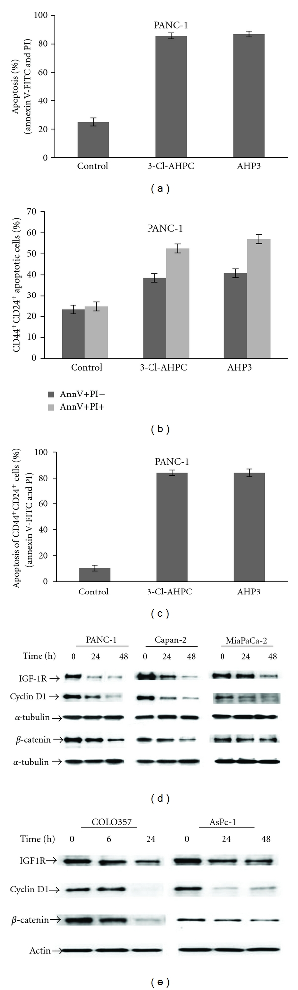
Pancreatic carcinoma has a dismal prognosis as it often presents as locally advanced or metastatic. We have found that exposure to adamantyl-substituted retinoid-related (ARR) compounds 3-Cl-AHPC and AHP3 resulted in growth inhibition and apoptosis induction in PANC-1, Capan-2, and MiaPaCa-2 pancreatic cancer cell lines. In addition, AHP3 and 3-Cl-AHPC inhibited growth and induced apoptosis in spheres derived from the CD44(+)/CD24(+) (CD133(+)/EpCAM(+)) stem-like cell population isolated from the pancreatic cancer cell lines. 3-Cl-AHPC-induced apoptosis was preceded by decreasing expression of IGF-1R, cyclin D1, β-catenin, and activated Notch-1 in the pancreatic cancer cell lines. Decreased IGF-1R expression inhibited PANC-1 proliferation, enhanced 3-Cl-AHPC-mediated apoptosis, and significantly decreased sphere formation. 3-Cl-AHPC inhibited the Wnt/β-catenin pathway as indicated by decreased β-catenin nuclear localization and inhibited Wnt/β-catenin activation of transcription factor TCF/LEF. Knockdown of β-catenin using sh-RNA also induced apoptosis and inhibited growth in pancreatic cancer cells. Thus, 3-Cl-AHPC and AHP3 induce apoptosis in pancreatic cancer cells and cancer stem-like cells and may serve as an important potential therapeutic agent in the treatment of pancreatic cancer.


Figures

Figure 1
Figure 1
3-Cl-AHPC- and AHP3-mediated proliferation inhibition and apoptosis induction in pancreatic cancer cell lines. The cells were exposed to 1 μM 3-Cl-AHPC or AHP3 for various times. (a) Proliferation inhibition was evaluated by MTT assay as described Section 2 and expressed as absorbance (OD) measured at 570 nm. The error bars represent the mean of three separate determinations ± the standard deviation (SD). (b) Induction of apoptosis in pancreatic cancer cells by 3-Cl-AHPC and AHP3. Cells were seeded at 1 × 10⁴ cells/mL and grown for 24 h and then exposed to 1 μM AHP3 or 3-Cl-AHPC for indicated times. Induction of apoptosis and cell death was assessed using Annexin V-FITC labeling with propidium iodide (PI) staining in COLO357, PANC-1, Capan-2 AsPc-1, or acridine orange/ethidium bromide staining in MiaPaCa-2. The error bars represent the mean of three separate determinations ± the standard deviation (SD). All treated samples are significantly different from vehicle control.
Figure 2
Figure 2
3-Cl-AHPC-mediated inhibition and degradation of pancreatic cancer stem cells spheres of CD133+, CD44+/CD24+/EpCAM+, and stem-like spheres of CD44+/CD24+ PANC-1 cells. ((a), (b) and (c), (d)) 3-Cl-AHPC and AHP3 exposure resulted in inhibition of CD44+/CD24+/EpCAM+ and CD133+cells growth and sphere formation and degradation of the derived spheres. ((e), (f)) AHP3 and 3-Cl-AHPC inhibited sphere formation and inhibition of growth and degradation of the CD44+/CD24+-derived spheres. For sphere formation, the CD44+/CD24+/EpCAM+, CD133+, and CD44+/CD24+ cells were sorted by flow cytometry and approximately 200–300 cells were seeded with B27 containing DMEM/F12 medium in 96-well low attachment plates and 1 μM 3-Cl-AHPC or AHP3 added either the day after seeding or 7 days following sphere formation. The sizes of spheres were photographed and measured on a 100 μm scale and magnification 400X using Olympus fluorescence microscope digital camera software and DP2-BSW software. The error bars represent the mean of 15 sphere determinations ± the standard deviation. ** was significantly different in comparison to control spheres. Data were analyzed by ANOVA, Tukey HSD test for multiple comparisons. **P < 0.01 versus control.
Figure 3
Figure 3
3-Cl-AHPC- (1 μM) and AHP3- (1 μM) mediated inhibition of CD44+/CD24+ stem-like cell sphere formation and degradation of spheres derived from MiaPaCa-2 and Capan-2 cell lines. 3-Cl-AHPC and AHP3 were added at the time cells were seeded ((a), (c)) or 7 days after cells sphere formation ((b), (d)). The ARR affect on sphere growth was assessed at days 7 and 14 (a), days 7 and 14 (b), days 14 and 21 (c), and days 14 and 21 (d), respectively. The error bars represent the mean of 15 sphere determinations ± the standard deviation. ** Was significantly different in comparison to control spheres. Data were analyzed by ANOVA, Tukey HSD test for multiple comparisons. **P < 0.01 versus control.
Figure 4
Figure 4
Dose-response effect of 3-Cl-AHPC on CD44+/CD24+ cells sphere formation and apoptosis in PANC-1 sphere cells. (a) Addition of 0.25, 0.5, and 1.0 μM 3-Cl-AHPC added at time of cell seeding inhibited sphere formation at 7 and 14 days. (b) 0.5 and 1.0 μM 3-Cl-AHPC inhibited sphere formation when added 7 days following sphere formation. (c) 1.0 μM 3-Cl-AHPC induced apoptosis in CD44+/CD24+ sphere cells as indicated by nuclear fragmentation detected by acridine orange/ethidium bromide and (d) DAPI staining. Spheres were visualized and photographed utilizing a fluorescence microscope. (e) Apoptosis of sphere cells as demonstrated by TUNEL assay. CD44+/CD24+ spheres were treated with 1.0 μM ARRs for 7 days (7D) after sphere formation. Details of slides preparation, visualization, antibodies utilized, and TUNNEL assay methodologies were as described in Section 2.
Figure 5
Figure 5
3-Cl-AHPC and AHP induced apoptosis in PANC-1 CD44+/CD24+ cells and 3-Cl-AHPC decreased expression of IGF-1R, cyclin D1, and β-catenin in pancreatic cancer cells. (a) Percentage of total apoptotic cells. (b) Percentage of CD44+/CD24+ cells in the early (Annexin V-FITC positive and PI negative) or late (Annexin V-FITC positive and PI positive) apoptotic cell populations. (c) Percentage of total CD44+/CD24+ apoptotic cells (Annexin V-FITC positive and PI positive). Cells were treated with 1.0 μM 3-Cl-AHPC and AHP3 for 96 h. Antibody-conjugated markers CD44-APC-Cy7, CD24-APC, Annexin V-FITC, and PI were used to detect apoptotic and CD44+/CD24+ cells from the same samples. The error bars represent the mean of three separate determinations ± the standard deviation. ((d), (e)) IGF-1R, cyclin D1, and β-catenin expression decreased following 3-Cl-AHPC exposure in pancreatic cancer cells.
Figure 6
Figure 6
3-Cl-AHPC decreased expression of IGF-1R, cyclin D1, β-catenin, and cleaved Notch-1 and increased levels of cleaved-caspase-3 in CD44+/CD24+spheres. (a) IGF-1R, cyclin D1, and β-catenin expression decreased cleaved-caspase-3 increased with no change in Notch-1 protein levels in spheres following exposure to 3-Cl-AHPC. Pancreatic cancer cells and PANC-1 spheres were exposed to 1.0 μM 3-Cl-AHPC for 7 days. Western blots were prepared as described in Materials and Methods. (b) mRNA expression of GLI1, GLI2, and Ptch1 in PANC-1 cells. Cells were grown in the presence of 1 μM 3-Cl-AHPC or vehicle alone (control). (c) Knockdown of IGF-1R expression by sh-IGF-1R inhibited sphere formation and enhanced ARR inhibition of sphere formation. The error bars represent the mean of three separate determinations +/− the standard deviation. •• was significantly different between spheres comprised of sh-vector cells treated with vehicle and 3-Cl-AHPC or AHP3. ♦♦ was significantly different between spheres comprised of sh-vector and IGF-1R-KD1 or IGF-1R-KD 2 at 7 and 14 days, respectively. ** was significantly different in comparison between IGF-1R-KDl/IGF-1R-KD2 spheres (vehicle treated) and IGF-1R-KD1/IGF-1R-KD2 spheres treated with 3-Cl-AHPC or AHP3. Data were analyzed by ANOVA, Tukey HSD test for multiple comparisons, ♦♦, ••, and **P < 0.01. (d) Knockdown (KD) of IGF-1R enhanced AHP3- and 3-Cl-AHPC-mediated apoptosis in the PANC-1 cells and IGF-IR protein expression in IGF-1R knockdown cells. Apoptosis was assessed using acridine orange/ethidium bromide staining as described in Section 2.
Figure 7
Figure 7
3-Cl-AHPC mediated inhibition of the activation of TCF/LEF in Wnt/β-catenin pathway and decreased of β-catenin nuclear localization. (a) 3-Cl-AHPC decreased nuclear β-catenin as indicated by Western blot using nuclear extracts and densitometric quantification. (b) Nuclear β-catenin in control- (i) and 3-Cl-AHPC- (ii) treated PANC-1 cells using confocal fluorescent microscope (magnification 40X). Cells were grown in eight chambered slides and then treated with 3-Cl-AHPC for 24 h. Slide was prepared as described in Section 2. DAPI was used for nuclear staining for 1 min and mounted the slide with prolong gold antifade kit. (c) 3-Cl-AHPC inhibited TCF/LEF activity in Wnt/β-catenin signaling in stably transfected Cignal TCF/LEF-Luc reporter PANC-1 cell lines and 50 mM LiCl was used as a positive control. For CD44/CD24 cells, TCF/LEF stably transfected cells were sorted by flow cytometry and followed the procedure same as wild type (Wt) stable cell line. Luciferase promoter activity values are expressed as fold using a total protein concentration for internal normalization. The error bars represent the mean of three separate determinations ± the standard deviation (SD). (d) 3-Cl-AHPC decreased Wnt/β-catenin signaling responsive c-Myc protein. ((e) and (f)) Knock down of β-catenin inhibited cell proliferations and enhanced more apoptosis in sh-β-catenin knockdown (KD) PANC-1 cell lines. Proliferation inhibition was evaluated after 72 h of seeding the cells by MTT assay and expressed as absorbance measured at 570 nm. The error bars represent the mean of three separate determinations ± the standard deviation (SD). ** (<0.01) was significantly different in comparison between sh-vector and Catenin-KDl/Catenin-KD2 and also between sh-vector and catenin-KD1/Catenin-KD2 treated with 3-Cl-AHPC, respectively.

References

    1. Bednar F, Simeone DM. Pancreatic cancer stem cells and relevance to cancer treatments. Journal of Cellular Biochemistry. 2009;107(1):40–45. - PubMed
    1. Clarke MF, Dick JE, Dirks PB, et al. Cancer stem cells—perspectives on current status and future directions: AACR workshop on cancer stem cells. Cancer Research. 2006;66(19):9339–9344. - PubMed
    1. Hermann PC, Huber SL, Herrler T, et al. Distinct populations of cancer stem cells determine tumor growth and metastatic activity in human pancreatic cancer. Cell Stem Cell. 2007;1(3):313–323. - PubMed
    1. Lee CJ, Dosch J, Simeone DM. Pancreatic cancer stem cells. Journal of Clinical Oncology. 2008;26(17):2806–2812. - PubMed
    1. Farhana L, Dawson MI, Xia Z, et al. Adamantyl-substituted retinoid-related molecules induce apoptosis in human acute myelogenous leukemia cells. Molecular Cancer Therapeutics. 2010;9(11):2903–2913. - PMC - PubMed