. 2012;7(5):e34142.
doi: 10.1371/journal.pone.0034142. Epub 2012 May 3.

Overexpression of the catalytically impaired Taspase1 T234V or Taspase1 D233A variants does not have a dominant negative effect in T(4;11) leukemia cells

Affiliations

Overexpression of the catalytically impaired Taspase1 T234V or Taspase1 D233A variants does not have a dominant negative effect in T(4;11) leukemia cells

Carolin Bier et al. PLoS One. 2012.

Abstract

Background: The chromosomal translocation t(4;11)(q21;q23) is associated with high-risk acute lymphoblastic leukemia of infants. The resulting AF4•MLL oncoprotein becomes activated by Taspase1 hydrolysis and is considered to promote oncogenic transcriptional activation. Hence, Taspase1's proteolytic activity is a critical step in AF4•MLL pathophysiology. The Taspase1 proenzyme is autoproteolytically processed in its subunits and is assumed to assemble into an αββα-heterodimer, the active protease. Therefore, we investigated here whether overexpression of catalytically inactive Taspase1 variants are able to interfere with the proteolytic activity of the wild type enzyme in AF4•MLL model systems.

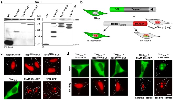
Methodology/findings: The consequences of overexpressing the catalytically dead Taspase1 mutant, Taspase1(T234V), or the highly attenuated variant, Taspase1(D233A), on Taspase1's processing of AF4•MLL and of other Taspase1 targets was analyzed in living cancer cells employing an optimized cell-based assay. Notably, even a nine-fold overexpression of the respective Taspase1 mutants neither inhibited Taspase1's cis- nor trans-cleavage activity in vivo. Likewise, enforced expression of the α- or β-subunits showed no trans-dominant effect against the ectopically or endogenously expressed enzyme. Notably, co-expression of the individual α- and β-subunits did not result in their assembly into an enzymatically active protease complex. Probing Taspase1 multimerization in living cells by a translocation-based protein interaction assay as well as by biochemical methods indicated that the inactive Taspase1 failed to assemble into stable heterocomplexes with the wild type enzyme.

Conclusions: Collectively, our results demonstrate that inefficient heterodimerization appears to be the mechanism by which inactive Taspase1 variants fail to inhibit wild type Taspase1's activity in trans. Our work favours strategies targeting Taspase1's catalytic activity rather than attempts to block the formation of active Taspase1 dimers to interfere with the pathobiological function of AF4•MLL.


Conflict of interest statement

Competing Interests: The authors have declared that no competing interests exist.

Figures

Figure 1
Figure 1. Analyzing Taspase1’s processing of AF4•MLL substrates in living cells.
A. Autoproteolysis of the Taspase1 proenzyme is assumed to trigger formation of the active αββα-heterodimer, which hydrolyses the AF4MLL fusion protein. Following processing, the cleavage products AF4MLL.N and MLL.C heterodimerize, forming a high molecular-weight protein complex resistant to degradation. Domain organization of the AF4MLL fusion. Taspase1 cleavage sites, S1 (QVDGADD) and S2 (QLDGVDD), are highlighted. NHD: N-terminal homology domain; ALF: AF4/LAF4/FMR2 homology domain; PHD: plant homeodomain; BrD: bromodomain; FRYN: F/Y rich domain N-terminal; TAD: transactivation domain; FRYC: F/Y rich domain C-terminal; SET: suppressor of variegation, enhancer of zeste and trithorax. Domains are not drawn to scale. B. Principle of the cell-based biosensor assay to analyze Taspase1-mediated AF4MLL processing. The indicator protein localizes predominantly to the cytoplasm but is continuously shuttling between the nucleus and the cytoplasm. Co-expression of active Taspase1 results in the proteolytic removal of the NES, thereby triggering nuclear accumulation of the green fluorescent indicator. C–D. Domains of the indicator protein, composed of GST, GFP, combinations of a nuclear import (?: NLS) and an export (?: NES) signal, combined with the indicated cleavage sites of AF4MLL. c. AM_S1/2 containing both cleavage sites is already partially processed by endogenous Taspase1 (left panel), but is completely nuclear upon expression of Taspase1-BFP (right panel). D. Indicator proteins containing only one cleavage site (AM_S1 or A M_S2) are cytoplasmic in their uncleaved state, whereas ectopic expression of active Taspase1 triggers their cleavage and complete nuclear accumulation. GFP/BFP were visualized by fluorescence microscopy in living HeLa transfectants 24 h after transfection. Scale bars, 10 µm. Dashed lines mark cytoplasmic/nuclear cell boundaries obtained from the corresponding phase contrast images.
Figure 2
Figure 2. Activity and complex formation of Taspase1 and catalytically inactive mutants. A.
Taspase1 processing of AF4MLL substrates in leukemic cells. Co-transfection of Tasp-GFP resulted in proteolytic cleavage and nuclear accumulation of the red fluorescent biosensor, AM_S2R, in K562 cells. In contrast, co-expression of TaspD233A-GFP leads to partial processing and nuclear translocation, while TaspT234V-GFP was completely inactive. Localization was analyzed 24 h post transfection. GFP/mCherry were visualized by fluorescence microscopy. Scale bars, 10 µm. B. Processing of AF4MLL substrates. Co-transfection of Tasp resulted in proteolytic cleavage of the biosensor AM_S2R in 293T cells as indicated by immunoblot. In contrast, TaspT234V was inactive in cis and trans. Proteins were visualized using α-GST or α-Taspase1 Abs. GapDH served as loading control. fl, unprocessed Taspase1; Taspβ, Taspase1 β-subunit.
Figure 3
Figure 3. Overexpression of inactive Taspase1 mutants does not inhibit Taspase1’s cis- or trans-cleavage activity.
A. Cells were transfected with 1 µg of AM_S2R, 0.1 µg Tasp-BFP together with the indicated amounts of inactive Taspase1 mutants or GFP expression plasmid, and analyzed 24 h later. Even co-transfection of a nine-fold excess of plasmids encoding the inactive Taspase1 variants did not affect AM_S2R processing in living HeLa cells. B. The number of HeLa (left panel) or leukemic K562 cells (right panel) showing cytoplasmic (C), cytoplasmic and nuclear (N/C) or nuclear (N) fluorescence was counted in at least 200 AM_S2R-expressing cells. Results from one representative experiment of each indicated cell line are shown. Whereas the number of cell displaying cytoplasmic fluorescence significantly decreased by trans-cleavage upon co-transfection of 0.1 µg Tasp-BFP expression plasmid (***: p<0.0001), no significant trans-dominant negative effect was evident for Taspase1 mutants. C. Taspase1 trans-cleavage of AM_S2R is unaffected by inactive Taspase1 mutants as shown by immunoblot analysis of 293T cells transfected with the indicated expression plasmids. Proteins and cleavage products were visualized using α-GST and α-Tasp Ab. GapDH served as loading control. D. Cis-cleavage of Taspase1 is not inhibited by inactive Taspase1 mutants as shown by immunoblot analysis of 293T cells transfected with 1 µg of the indicated expression plasmids.
Figure 4
Figure 4. Probing Taspase1 multimerization in living cells.
A. Heterocomplex formation of Taspase1 and Taspase1 variants shown by co-immunoprecipitation (IP). IPs of 293T cell extracts co-transfected with the indicated expression constructs were carried out using α-GFP Ab-coated magnetic beads and μ-MACS columns. Precipitated proteins were identified by immunoblot using the indicated antibodies. Input: Total amount of cell lysate. IP: immunoprecipitated proteins. *: GFP-degradation products . B. Principle of the translocation based protein-protein interaction assay. The TaspCyt fusion is composed of GFP, Taspase1 and a NES (?) and thus, continuously shuttling between the nucleus and the cytoplasm. The red-fluorescent Taspase1 variants (Tasp-mCherry prey) accumulate at the nucleus/nucleolus. Upon efficient protein-protein interaction, the GFP-tagged cytoplasmic TaspCyt co-localizes with the Tasp-mCherry prey to the nucleus/nucleolus in living cells. C. Localization of indicated proteins in the absence of potential interaction partners. D. Neither co-expression of WT nor inactive Taspase1 variants resulted in strong nuclear/nucleolar translocation of TaspCyt. Co-expression of NPM1-RFP, known to strongly interact with Taspase1, triggered nuclear/nucleolar translocation of TaspCyt (positive control). In contrast, co-expression of the non-interacting nucleolar RevM10BL-RFP protein showed no effect (negative control) as visualized by fluorescence microscopy in living HeLa transfectants. Scale bars, 10 µm.
Figure 5
Figure 5. Models illustrating how Taspase1 heterocomplex formation determines the biological effects of overexpressing inactive Taspase1 mutants. A–C:
Heterodimer model - allowing inhibition of Taspase1 function by trans dominant mutants. A. Upon translation, the Taspase1 zymogen dimerizes and following autoproteolysis matures into an asymmetric Taspase1αββα-heterodimer, representing the active protease. Taspase1 exist in equilibrium of unprocessed Taspase1 monomers, unprocessed Taspase1 dimers, and active processed Taspase1αββα-heterodimers. The Taspase1αββα-heterodimers may further dissociate into free Taspase1α and Taspase1β subunits. B. Co-expression of an excess of inactive Taspase1 variants results in the formation of catalytically impaired heterodimers, reducing the concentration of active Taspase1 molecules. C. Consequently, AF4MLL processing is inhibited allowing its degradation by SIAH1/2, thereby preventing the activation of cellular proliferation programs. D–F: Monomer model - predicting Taspase1’s resistance to enforced expression of inactive mutants. D. The Taspase1αβ proenzyme is autoproteolytically cleaved, forming an active Taspase1αβ monomer. The processed Taspase1αβ monomer seems to exist also as a Taspase1αββα-heterodimer, and potentially in equilibrium with its subunits. E. Overexpression of inactive Taspase1 variants does not affect the concentration and activity of Taspase1αβ monomers. F. Hence, Taspase1αβ monomers are able to cleave the AF4MLL fusion protein, resulting in the formation of a SIAH-resistant AF4MLL complex allowing the activation of target genes driving oncogenesis.

References

    1. Meyer C, Kowarz E, Hofmann J, Renneville A, Zuna J, et al. New insights to the MLL recombinome of acute leukemias. Leukemia. 2009;23:1490–1499. - PubMed
    1. Montes R, Ayllon V, Gutierrez-Aranda I, Prat I, Hernandez-Lamas MC, et al. Enforced expression of MLL-AF4 fusion in cord blood CD34+ cells enhances the hematopoietic repopulating cell function and clonogenic potential but is not sufficient to initiate leukemia. Blood. 2011;117:4746–4758. - PubMed
    1. Bursen A, Schwabe K, Ruster B, Henschler R, Ruthardt M, et al. The AF4.MLL fusion protein is capable of inducing ALL in mice without requirement of MLL.AF4. Blood. 2010;115:3570–3579. - PubMed
    1. Hsieh JJ, Ernst P, Erdjument-Bromage H, Tempst P, Korsmeyer SJ. Proteolytic cleavage of MLL generates a complex of N- and C-terminal fragments that confers protein stability and subnuclear localization. Mol Cell Biol. 2003;23:186–194. - PMC - PubMed
    1. Yokoyama A, Kitabayashi I, Ayton PM, Cleary ML, Ohki M. Leukemia proto-oncoprotein MLL is proteolytically processed into 2 fragments with opposite transcriptional properties. Blood. 2002;100:3710–3718. - PubMed

Publication types

MeSH terms

LinkOut - more resources