. 2012;7(5):e36402.
doi: 10.1371/journal.pone.0036402. Epub 2012 May 3.

Active PI3K pathway causes an invasive phenotype which can be reversed or promoted by blocking the pathway at divergent nodes

Affiliations

Active PI3K pathway causes an invasive phenotype which can be reversed or promoted by blocking the pathway at divergent nodes

Jeffrey J Wallin et al. PLoS One. 2012.

Abstract

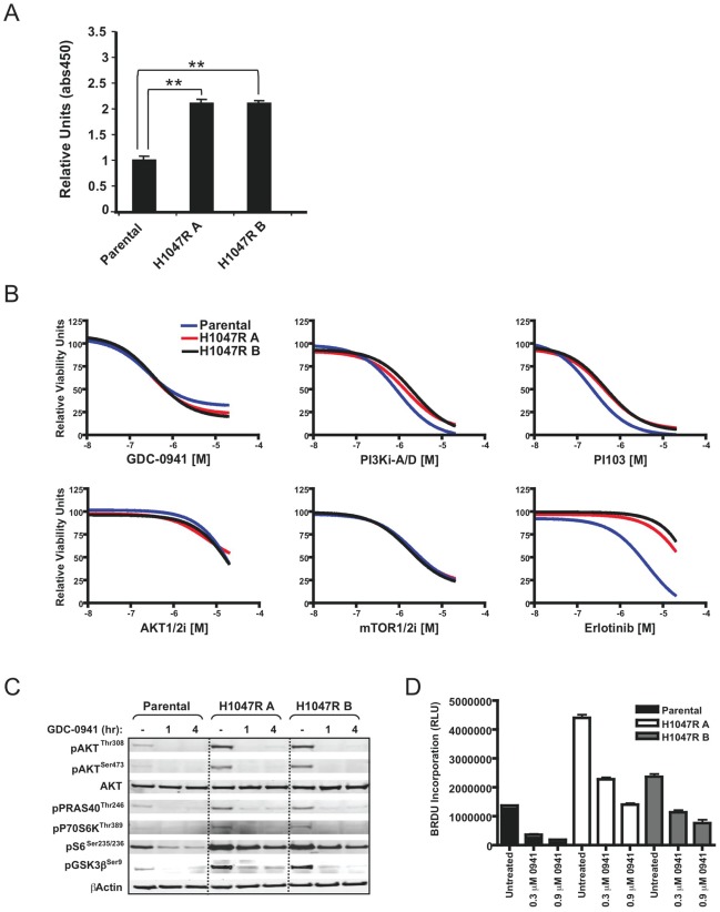
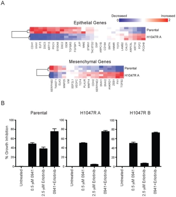
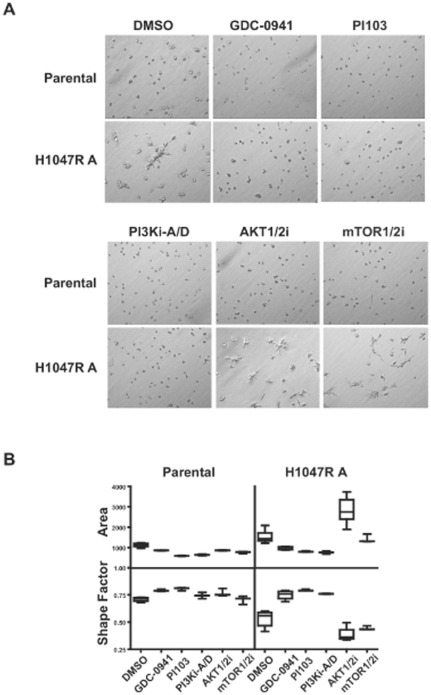
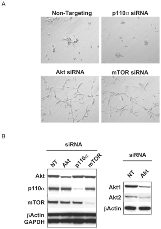
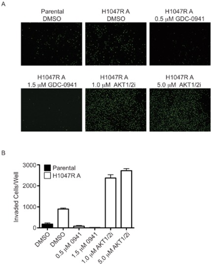
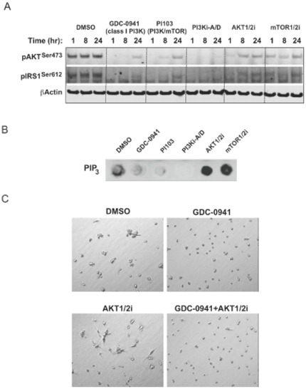
The PTEN/PI3K pathway is commonly mutated in cancer and therefore represents an attractive target for therapeutic intervention. To investigate the primary phenotypes mediated by increased pathway signaling in a clean, patient-relevant context, an activating PIK3CA mutation (H1047R) was knocked-in to an endogenous allele of the MCF10A non-tumorigenic human breast epithelial cell line. Introduction of an endogenously mutated PIK3CA allele resulted in a marked epithelial-mesenchymal transition (EMT) and invasive phenotype, compared to isogenic wild-type cells. The invasive phenotype was linked to enhanced PIP(3) production via a S6K-IRS positive feedback mechanism. Moreover, potent and selective inhibitors of PI3K were highly effective in reversing this phenotype, which is optimally revealed in 3-dimensional cell culture. In contrast, inhibition of Akt or mTOR exacerbated the invasive phenotype. Our results suggest that invasion is a core phenotype mediated by increased PTEN/PI3K pathway activity and that therapeutic agents targeting different nodes of the PI3K pathway may have dramatic differences in their ability to reverse or promote cancer metastasis.


Conflict of interest statement

Competing Interests: The authors have read the journal's policy and have the following conflicts: J. Wallin, J. Guan, K. Edgar, A. Torres, P. Haverty, J. Eastham-Anderson, W. Zhou, G. Francis, K. Hoeflich, M. Belvin and L. Friedman are employed at Genentech, Inc. S. Griffin, J. Goodall, K. Grimshaw and C. Torrance are employed at Horizon Discovery Ltd. This does not alter the authors' adherence to all the PLoS ONE policies on sharing data and materials.

Figures

Figure 1
Figure 1. Effects of the H1047R mutation on PI3K pathway signaling, cell viability and proliferation.
(A) Analysis of PIP3 levels in parental and knock-in clones. (B) Parental and knock-in clones were cultured with dose-titrated small molecule inhibitors and cell viability was assessed after four days. (C) Parental and knock-in clones were cultured in the presence or absence of GDC-0941 dosed at an EC50 concentration for the time points indicated and analyzed by Western blotting. (D) The parental and knock-in clones were cultured in the presence or absence of GDC-0941 for 48 hours and proliferation was assessed in triplicate samples by BrdU labeling.
Figure 2
Figure 2. MCF10A cells undergo EMT-like changes in the presence of a PI3K mutation.
(A) Comparative gene expression analysis of the parental and knock-in clone. Expression profile differences of commonly associated epithelial and mesenchymal genes by microarray are shown by heat map for a ratio of gene expression levels (H1047R A to parental). (B) Effects of GDC-0941 and erlotinib combination treatment in parental and knock-in cell lines. Percent growth inhibition data from a viability assay is shown at EC50 doses of single agent GDC-0941 and 2.5 µM erlotinib after drug incubation. Each bar indicates mean % inhibition ±SEM from quadruplicate wells.
Figure 3
Figure 3. MCF10A knock-in cells show a more invasive phenotype in 3-D cell culture.
(A) Parental and H1047R A cells were cultured for 2 days in the presence or absence of GDC-0941 (0.5 µM), PI103 (0.5 µM), PI3Ki-A/D (2 µM), AKT1/2i (5 µM) or mTOR1/2i (5 µM). (B) A mathematical distribution of acinar size (area) and shape (shape factor) was used to assess morphology changes with drug treatments on day 2. Data are plotted as the mean (horizontal line), middle 50% of data (box), and 95% confidence interval (lines). Pair-wise comparisons to the DMSO control were done by Student's t test. GDC-0941 and PI103 treatments resulted in significant morphology changes in both the parental and H1047R A clone (p<0.02, area or shape factor). PI3Ki-A/D treatment resulted in significant morphological changes in parental (p<0.006, area) or the H1047R clone (p<0.003, area or shape factor). Statistical significance was also achieved in the H1047R clone with the AKT1/2i (p<0.0002, area or shape factor) or mTOR1/2i (p = 0.03, shape factor).
Figure 4
Figure 4. siRNA knockdown of p110α, Akt or mTOR phenocopies small molecule inhibitor treatments.
(A) Knock-in cells (H1047R A) were transfected with the indicated siRNAs and plated in the 3-D culture assay for 5 days. (B) Assessment of siRNA knockdown by Western blot in the knock-in clone (H1047R A) 48 hours after transfection (NT = Non-targeting control siRNA).
Figure 5
Figure 5. MCF10A knock-in cells show enhanced migration through matrigel.
(A) Cells were plated under the conditions indicated and allowed to migrate for 24 hours. Representative images for each condition are shown. (B) Quantification of migrated cells in triplicate wells.
Figure 6
Figure 6. PI3K signaling responses to PI3K pathway inhibition.
(A) Knock-in cells (H1047R A) were cultured with inhibitors at concentrations used in the 3-D culture assay and analyzed by Western blotting for indicated treatment times. (B) PIP3 levels assessed at 48 hours post treatment with 3-D culture concentrations of inhibitors in the knock-in clone (H1047R A). (C) PI3K and Akt inhibitor effects can be distinguished in 3-D culture through variations in cellular morphology. Phase contrast images of the knock-in clone (H1047R A) show representative phenotypes of treatment with 0.5 µM GDC-0941, 5 µM AKT1/2i, or the same compound doses in combination after 2 days in culture.

Similar articles

Cited by

References

    1. Osaki M, Oshimura M, Ito H. PI3K-Akt pathway: its functions and alterations in human cancer. Apoptosis. 2004;9:667–676. - PubMed
    1. Kang S, Bader AG, Vogt PK. Phosphatidylinositol 3-kinase mutations identified in human cancer are oncogenic. Proc Natl Acad Sci U S A. 2005;102:802–807. - PMC - PubMed
    1. Samuels Y, Wang Z, Bardelli A, Silliman N, Ptak J, et al. High frequency of mutations of the PIK3CA gene in human cancers. Science. 2004;304:554. - PubMed
    1. Zhang S, Yu D. PI(3)king apart PTEN's role in cancer. Clin Cancer Res. 2010;16:4325–4330. - PubMed
    1. Haruta T, Uno T, Kawahara J, Takano A, Egawa K, et al. A rapamycin-sensitive pathway down-regulates insulin signaling via phosphorylation and proteasomal degradation of insulin receptor substrate-1. Mol Endocrinol. 2000;14:783–794. - PubMed

MeSH terms

Substances