. 2012;7(5):e36935.
doi: 10.1371/journal.pone.0036935. Epub 2012 May 3.

Role of stem cells in human uterine leiomyoma growth

Affiliations

Role of stem cells in human uterine leiomyoma growth

Masanori Ono et al. PLoS One. 2012.

Abstract

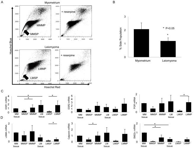
Background: Uterine leiomyoma is the most common benign tumor in reproductive-age women. Each leiomyoma is thought to be a benign monoclonal tumor arising from a single transformed myometrial smooth muscle cell; however, it is not known what leiomyoma cell type is responsible for tumor growth. Thus, we tested the hypothesis that a distinct stem/reservoir cell-enriched population, designated as the leiomyoma-derived side population (LMSP), is responsible for cell proliferation and tumor growth.

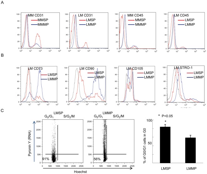
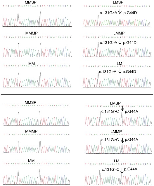
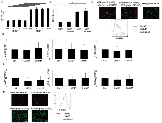
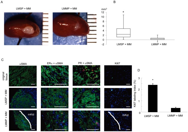
Principal findings: LMSP comprised approximately 1% of all leiomyoma and 2% of all myometrium-derived cells. All LMSP and leiomyoma-derived main population (LMMP) but none of the side or main population cells isolated from adjacent myometrium carried a mediator complex subunit 12 mutation, a genetic marker of neoplastic transformation. Messenger RNA levels for estrogen receptor-α, progesterone receptor and smooth muscle cell markers were barely detectable and significantly lower in the LMSP compared with the LMMP. LMSP alone did not attach or survive in monolayer culture in the presence or absence of estradiol and progestin, whereas LMMP readily grew under these conditions. LMSP did attach and survive when directly mixed with unsorted myometrial cells in monolayer culture. After resorting and reculturing, LMSP gained full potential of proliferation. Intriguingly, xenografts comprised of LMSP and unsorted myometrial smooth muscle cells grew into relatively large tumors (3.67 ± 1.07 mm(3)), whereas xenografts comprised of LMMP and unsorted myometrial smooth muscle cells produced smaller tumors (0.54 ± 0.20 mm(3), p<0.05, n = 10 paired patient samples). LMSP xenografts displayed significantly higher proliferative activity compared with LMMP xenografts (p<0.05).

Conclusions: Our data suggest that LMSP, which have stem/reservoir cell characteristics, are necessary for in vivo growth of leiomyoma xenograft tumors. Lower estrogen and progesterone receptor levels in LMSP suggests an indirect paracrine effect of steroid hormones on stem cells via the mature neighboring cells.


Conflict of interest statement

Competing Interests: The authors have declared that no competing interests exist.

Figures

Figure 1
Figure 1. Isolation and characterization of human MMSP and LMSP.
(A) (Left upper) Distribution of the SP and MP cells within all Hoechst 33342-stained living cells isolated from human myometrium. (Right upper) Addition of 50 µM reserpine resulted in the disappearance of the MMSP fraction. (Left lower) Distribution of SP and MP cells isolated from human leiomyoma. (Right lower) Addition of 50 µM reserpine resulted in the disappearance of the LMSP fraction. (B) Average % SP of normal myometrium and leiomyomas from 10 different patients are shown (2.07%±0.46 vs. 1.19%±0.23, P<0.05). Error bars represent SEM. (C) mRNA expression of ovarian steroid receptors and (D) smooth muscle cell markers was examined by real-time RT-PCR and normalized for GAPDH expression. Each bar indicates the mean ± SEM of the relative expression obtained from three independent experiments using three individual samples. *, P<0.05.
Figure 2
Figure 2. Cell surface marker antigens in LMSP.
(A) Expression patterns of endothelial cell surface markers (CD31, Platelet Endothelial Cell Adhesion Molecule-1) and hematopoietic (CD45, Leukocyte common antigen) in MMSP, LMSP (red); and MMMP, LMMP (blue). In LMSP, most of the cells were negative for endothelial and hematopoietic cell markers. (B) Expression patterns of bone marrow mesenchymal stem cell surface markers (CD73, CD90, CD105, STRO-1) in MMSP, LMSP (red); and MMMP, LMMP (blue). These bone mesenchymal cell surface antigens were not able to identify LMSP. Mouse FITC-labeled IgG1 (BD Biosciences) was used as an isotypic control for staining of total myometrial or leiomyoma cells (black). (C) Cell cycle status of LMSP and LMMP was determined by Hoechst 33342 and Pyronin Y staining. The left lower quadrant corresponds to the G0 phase. Flow cytometry analysis revealed that 83.67%±5.73 of LMSP but only 58.50%±6.06 of LMMP were in the G0 phase.
Figure 3
Figure 3. Sequence chromatograms showing somatic mutations in MED12 codon 44 in LMSP.
Examples of genomic DNA sequencing traces in codon 44-mutated samples are shown. Mutated bases are indicated by arrows.
Figure 4
Figure 4. Cell culture of LMSP.
(A) Effects of E2 and/or P on the viability of LMSP as determined by the MTS assay. Each bar indicates the mean ± SEM of the absorbance at 490 nm obtained from three independent experiments using three individual samples. *, P<0.05. (B) Effects of indirect co-culturing with MM on the viability of LMSP. *, P<0.05. (C) Side or main populations in mixed co-cultures with MM at a 1∶1 ratio were identified after initially dye-labeling each cell type. Cells originating from LMSP, LMMP, and MM in mixed co-cultures were identified using long-lasting status PKH-26 (red) or PKH-67 (green). Scale bars, 100 µm. (D) mRNA expression of the ovarian steroid receptors and (E) smooth muscle cell markers was examined by real-time RT-PCR and normalized for GAPDH expression. Each bar indicates the mean ± SEM of the relative expression obtained from three independent experiments using three individual samples. *, P<0.05. (F) Expression of αSMA protein in LMSP and LMMP after culturing. Scale bars, 50 µm (upper), 100 µm (lower).
Figure 5
Figure 5. Generation of leiomyoma tumors from LMSP.
(A) Macroscopic visualization of the transplanted site 12 weeks after xenotransplantation. (B) Xenografts were analyzed in terms of tumor volume. Each bar indicates the mean ± SEM. *, P<0.05, n = 10 paired patient samples. (C) Original tissue and generated tumor were analyzed by immunohistochemistry. Nuclei were stained with DAPI (blue). Scale bars, 100 µm. D, Ki 67 labeling index was calculated in transplanted tumors.

References

    1. Walker CL, Stewart EA. Uterine fibroids: the elephant in the room. Science. 2005;308:1589–1592. - PubMed
    1. Okolo S. Incidence, aetiology and epidemiology of uterine fibroids. Best Pract Res Clin Obstet Gynaecol. 2008;22:571–588. - PubMed
    1. Wallach EE, Vlahos NF. Uterine myomas: an overview of development, clinical features, and management. Obstet Gynecol. 2004;104:393–406. - PubMed
    1. Stewart EA. Uterine fibroids. Lancet. 2001;357:293–298. - PubMed
    1. Szotek PP, Chang HL, Zhang L, Preffer F, Dombkowski D, et al. Adult mouse myometrial label-retaining cells divide in response to gonadotropin stimulation. Stem Cells. 2007;25:1317–1325. - PubMed

Publication types

MeSH terms