. 2011 Dec;23(4):185-96.
doi: 10.5792/ksrr.2011.23.4.185. Epub 2011 Nov 30.

Management of focal chondral lesion in the knee joint

Affiliations

Management of focal chondral lesion in the knee joint

Seung-Suk Seo et al. Knee Surg Relat Res. 2011 Dec.

Abstract

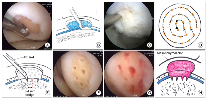
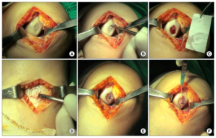
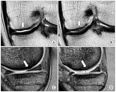
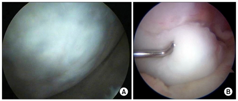
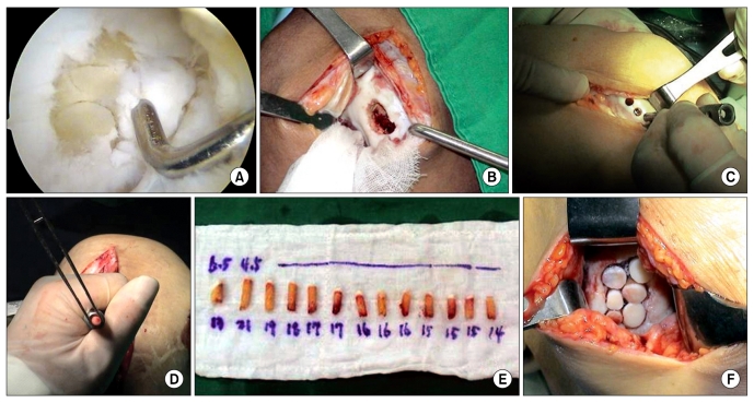
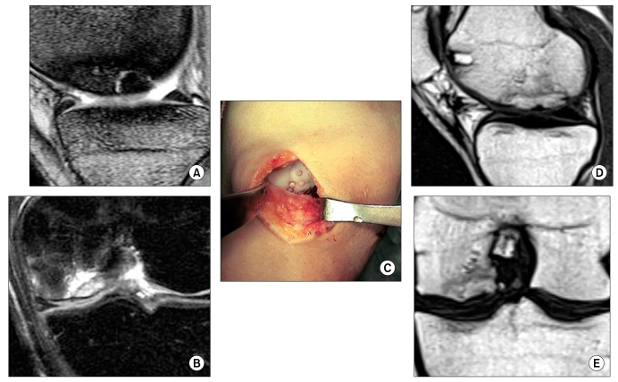
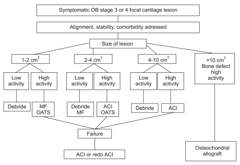
Articular cartilage does not contain vascular, nervous and lymphatic tissue and chondrocytes hardly participate in the healing or repair process of chondral tissue because of being surrounded by plenty of extracellular matrix. Therefore, the injury to articular cartilage frequently requires an operative treatment. The goal of surgical repair of articular cartilage is to regenerate nearly normal chondral tissue and prevent degenerative arthritis caused by the articular cartilage defect. Microfracture is a kind of cartilage repair procedure that makes a fibrin clot containing mesenchymal stem cells in the chondral lesion. Microfracture is a simple procedure but it has a disadvantage that the repaired tissue is fibrocartilage. Autologous chondrocyte implantation has an advantage that it implants fully differentiated chondrocytes to the lesion, which theoretically produces hyaline cartilage. Its disadvantages are that it is a two stage and a costly procedure. Osteochondral autograft transplantation is a one stage procedure and repairs the lesion with hyaline cartilage. But its limitation is the lack of donor site availability. Surgeons who understand the theoretical background, indications, surgical methods, rehabilitation, complications, and clinical course of cartilage repair procedures can achieve the goal of preventing degenerative arthritis.

Keywords: Articular cartilage; Focal chondral lesion; Knee joint; Management.


Figures

Fig. 1
Fig. 1
Surgical procedure of microfracture. (A) Unstable cartilage flap and calcified cartilage bed is debrided with open curette. (B) It is important to debride the calcified cartilage layer and make a well-contained pocket surrounded healthy cartilage (well-shouldered). (C) Subchondral bone is punctured with an awl. (D) Microfracture is circumferentially performed from periphery to center. (E) The penetration of subchondral bone is 3 to 4 mm deep and apart. (F) Arthroscopic photograph showing the final step of microfracture. (G) Mesenchymal blood egress from bone marrow through subchondral holes. (H) It is important for tissue regeneration to keep the mesenchymal clot in the defect.
Fig. 2
Fig. 2
Surgical procedure of the 1st generation autologous chondrocyte implantation. (A) Outerbridge 4 lesion in the medial femoral condyle. (B) Debridement of the calcified cartilage layer and unstable chondral flap. (C) Defect size is measured with a sterile paper. A 2 mm oversized template is needed. (D) Periosteal flap is excised from the proximal medial tibia. (E) The periosteal flap watertightly covers the defect. (F) Chondrocyte suspension is injected to the defect through a plastic 18-gauge angiocath needle.
Fig. 3
Fig. 3
MRI findings before and after autologous chondrocyte implantation (ACI). (A) Coronal proton density image showing the focal chondral defect with thinning of the cartilage. (B) Coronal proton density image showing the slightly hyperintense repaired tissue at 26 months after ACI. (C) Sagittal T2 fast spin echo (FSE) image showing the thinning of the cartilage and irregular elevation of the subchondral bone plate. (D) Sagittal T2 FSE image showing the hypertrophied regenerated cartilage tissue with focal hyperintense area at 26 months after ACI.
Fig. 4
Fig. 4
Second look arthroscopic findings after autologous chondrocyte implantation (ACI). (A) Regenerated hyaline like tissue after ACI, of which surface shows a smooth and well-incorporated margin. (B) Graft hypertrophy after 1st generation ACI, which causes a locking of the knee.
Fig. 5
Fig. 5
Surgical procedure of osteochondral autograft transplantation. (A) Arthroscopic determination of the number and size of grafts needed after debridement of cartilage lesion. (B) Open procedure. (C) Harvesting the osteochondral plug with a tubular chisel from the lateral supracondylar ridge. The tubular chisel must be perpendicularly located to the chondral surface. (D) Introduction of the osteochondral plug through a drill guide. (E) Harvested osteochondral plugs. Size and length of plugs are marked on the wet gauze. (F) Focal cartilage defect reconstructed with multiple osteochondral plugs. It is important to make a congruent surface with the adjacent cartilage.
Fig. 6
Fig. 6
Osteochondral plug fixation for the treatment of osteochondritis dissecans (OCD). (A) Sagittal T2 fast spin echo (FSE) image showing stage 3 OCD lesion in the medial femoral condyle. The lesion is partially separated. (B) Coronal T2 fat suppression FSE image showing OCD lesion with focal bone marrow edema. (C) Fixation of OCD with multiple osteochondral plugs. (D) Sagittal image. (E) Coronal image showing the OCD lesions completely incorporated to the host bone at 24 months after surgery.
Fig. 7
Fig. 7
Treatment algorithm for focal chondral lesions. Before treatment, it is important to assess the presence of correctable lesions (alignment, stability etc.). The treatment decision is guided by the size of the defect and the patient's demands. OB: outerbridge, MF: microfracture, OAT: osteochondral autograft transplantation, ACI: autologous chondrocyte implantation.

References

    1. Buckwalter JA, Mankin HJ. Articular cartilage. Part II: degeneration and osteoarthrosis, repair, regeneration, and transplantation. J Bone Joint Surg Am. 1997;79:612–632.
    1. Shah MR, Kaplan KM, Meislin RJ, Bosco JA., 3rd Articular cartilage restoration of the knee. Bull NYU Hosp Jt Dis. 2007;65:51–60. - PubMed
    1. Curl WW, Krome J, Gordon ES, Rushing J, Smith BP, Poehling GG. Cartilage injuries: a review of 31,516 knee arthroscopies. Arthroscopy. 1997;13:456–460. - PubMed
    1. Craig W, David JW, Ming HZ. A current review on the biology and treatment of the articular cartilage defects (part I & part II) J Musculoskelet Res. 2003;7:157–181.
    1. Messner K, Maletius W. The long-term prognosis for severe damage to weight-bearing cartilage in the knee: a 14-year clinical and radiographic follow-up in 28 young athletes. Acta Orthop Scand. 1996;67:165–168. - PubMed

LinkOut - more resources