

Simultaneous bilateral quadriceps tendon rupture in patient with chronic renal failure
- PMID: 22570843
- PMCID: PMC3341810
- DOI: 10.5792/ksrr.2011.23.4.244
Simultaneous bilateral quadriceps tendon rupture in patient with chronic renal failure
Abstract
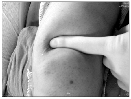
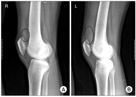
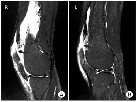
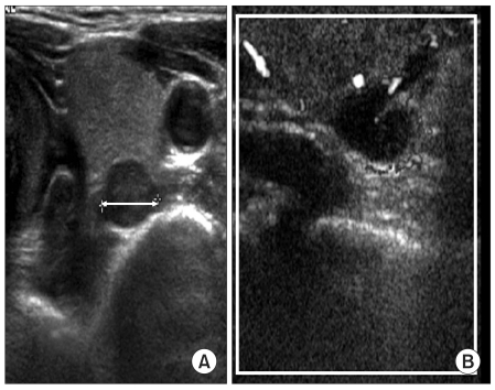
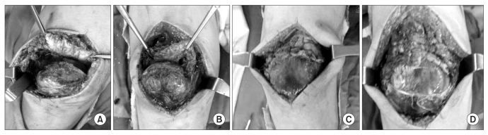
Simultaneous bilateral spontaneous rupture of the quadriceps tendon is a very rare condition and only a few cases have been reported in the literature. The etiology is not clear yet. But it occurs infrequently in patients with chronic metabolic disorders. A 30-year-old female patient with simultaneous bilateral spontaneous quadriceps tendon rupture visited our hospital. She had chronic renal failure and her parathyroid hormone level was elevated due to parathyroid adenoma. We report a surgical repair of both quadriceps tendons of a patient with chronic renal failure as well as management of hyperparathyroidism.
Keywords: End-stage renal failure; Parathyroid hormone; Quadriceps tendon.
Figures
References
-
- Steiner CA, Palmer LH. Simultaneous bilateral rupture of the quadriceps tendon. Am J Surg. 1949;78:752–755. - PubMed
-
- De Franco P, Varghese J, Brown WW, Bastani B. Secondary hyperparathyroidism, and not beta 2-microglobulin amyloid, as a cause of spontaneous tendon rupture in patients on chronic hemodialysis. Am J Kidney Dis. 1994;24:951–955. - PubMed
-
- Shiota E, Tsuchiya K, Yamaoka K, Kawano O. Spontaneous major tendon ruptures in patients receiving long-term hemodialysis. Clin Orthop Relat Res. 2002;(394):236–242. - PubMed
-
- Shah MK. Simultaneous bilateral rupture of quadriceps tendons: analysis of risk factors and associations. South Med J. 2002;95:860–866. - PubMed
-
- Preston FS, Adicoff A. Hyperparathyroidism with avulsion of three major tendons. Report of a case. N Engl J Med. 1962;266:968–971. - PubMed
LinkOut - more resources
Full Text Sources