. 2012 Jul;10(7):958-68.
doi: 10.1158/1541-7786.MCR-11-0616. Epub 2012 May 9.

Cdc42 and the guanine nucleotide exchange factors Ect2 and trio mediate Fn14-induced migration and invasion of glioblastoma cells

Affiliations

Cdc42 and the guanine nucleotide exchange factors Ect2 and trio mediate Fn14-induced migration and invasion of glioblastoma cells

Shannon P Fortin et al. Mol Cancer Res. 2012 Jul.

Abstract

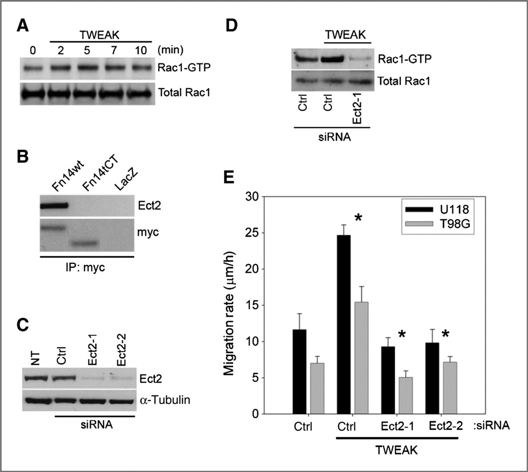
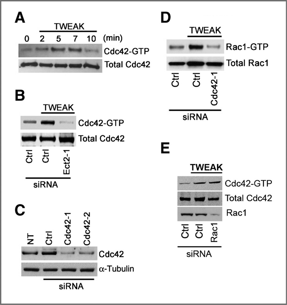
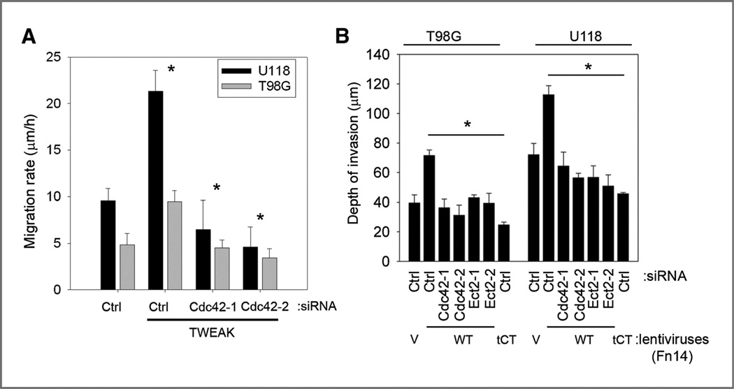
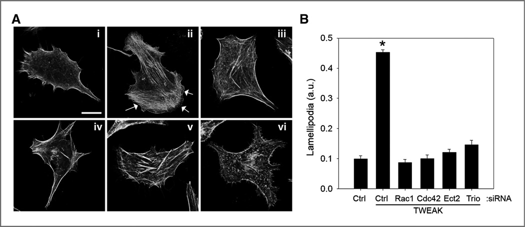
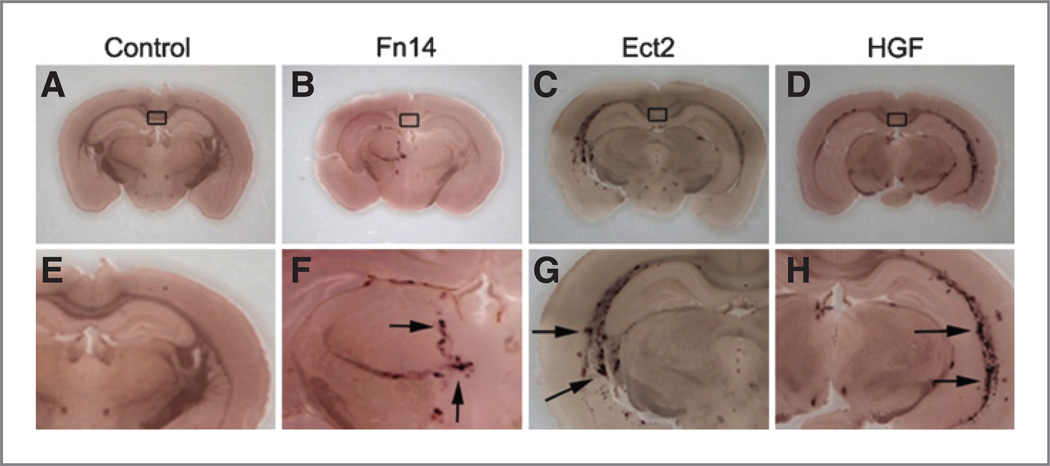
Malignant glioblastomas are characterized by their ability to infiltrate into normal brain. We previously reported that binding of the multifunctional cytokine TNF-like weak inducer of apoptosis (TWEAK) to its receptor fibroblast growth factor-inducible 14 (Fn14) induces glioblastoma cell invasion via Rac1 activation. Here, we show that Cdc42 plays an essential role in Fn14-mediated activation of Rac1. TWEAK-treated glioma cells display an increased activation of Cdc42, and depletion of Cdc42 using siRNA abolishes TWEAK-induced Rac1 activation and abrogates glioma cell migration and invasion. In contrast, Rac1 depletion does not affect Cdc42 activation by Fn14, showing that Cdc42 mediates TWEAK-stimulated Rac1 activation. Furthermore, we identified two guanine nucleotide exchange factors (GEF), Ect2 and Trio, involved in TWEAK-induced activation of Cdc42 and Rac1, respectively. Depletion of Ect2 abrogates both TWEAK-induced Cdc42 and Rac1 activation, as well as subsequent TWEAK-Fn14-directed glioma cell migration and invasion. In contrast, Trio depletion inhibits TWEAK-induced Rac1 activation but not TWEAK-induced Cdc42 activation. Finally, inappropriate expression of Fn14 or Ect2 in mouse astrocytes in vivo using an RCAS vector system for glial-specific gene transfer in G-tva transgenic mice induces astrocyte migration within the brain, corroborating the in vitro importance of the TWEAK-Fn14 signaling cascade in glioblastoma invasion. Our results suggest that the TWEAK-Fn14 signaling axis stimulates glioma cell migration and invasion through two GEF-GTPase signaling units, Ect2-Cdc42 and Trio-Rac1. Components of the Fn14-Rho GEF-Rho GTPase signaling pathway present innovative drug targets for glioma therapy.


Conflict of interest statement

Disclosure of Potential Conflicts of Interest

No potential conflicts of interests were disclosed.

Figures

Figure 1
Figure 1
Ect2 binds to Fn14 and regulates TWEAK-induced Rac1 activation and glioma cell migration. A, T98G cells were cultured in reduced serum followed by treatment with 100 ng/mL TWEAK for the indicated times. Whole-cell lysates were assessed for active Rac1 via affinity pull-down with the Pak1-binding domain (PBD). B, T98G cells were infected with adenoviruses expressing myc-tagged Fn14 wild-type (wt), Fn14 C-terminal–truncated (Fn14tCT), or control LacZ. Whole-cell lysates were immunoprecipitated (IP) for myc with subsequent Western blot analysis for Ect2 and myc. C, T98G cells were transfected with 2 siRNA oligonucleotides targeting Ect2 and assessed for the level of shutdown efficiency relative to nontransfected (NT) or ctrl siRNA transfection targeting nonmammalian luciferase (Ctrl) by Western blot analysis. D, T98G cells were cultured in reduced serum followed by treatment with TWEAK (100 ng/mL) for 10 minutes in the presence of either control (ctrl) luciferase–targeted siRNA or siRNA-targeting Ect2 and were assessed by Western blotting. E, glioma cells were transfected with siRNA targeting either control (ctrl) luciferase or 2 independent siRNA oligonucleotides to Ect2 (Ect2-1 and Ect2-2). After 24 hours, cells were cultured in reduced serum medium (0.5% FBS) for 16 hours and were seeded onto 10-well glass slides precoated with 10 µg/mL human laminin. Cells were either left untreated or treated with TWEAK, and glioma cell migration was assessed over 24 hours. Data represent the average of 3 independent experiments (*, P < 0.01).
Figure 2
Figure 2
TWEAK stimulation of Rac1 activation is dependent upon Cdc42. A, T98G cells were treated with TWEAK for the indicated times and assayed for Cdc42 activity using the Pak1-binding domain (PBD) assay. B, T98G cells were transfected with Ect2 siRNA oligonucleotides or luciferase control siRNA (Ctrl). Cells were then cultured under reduced serum (0.5% FBS) for 24 hours, followed by treatment with TWEAK for 5 minutes and then assessed for Cdc42 activity. C, T98G cells were either left nontransfected (NT), transfected with 2 independent siRNAs designed against Cdc42 (Cdc42-1 and Cdc42-2), or transfected with control siRNA. Cells were then lysed, and Western blot analysis for Cdc42 was conducted to confirm protein depletion. D, cells transfected with either control or Cdc42-targeting siRNA were cultured under reduced serum for 16 hours. Cells were then either left untreated or treated with TWEAK for 10 minutes and assayed for Rac1 activity. E, cells transfected with either control or Rac1-targeting siRNA were cultured under reduced serum for 16 hours. Cells were then either left untreated or treated with TWEAK for 5 minutes and assessed for Cdc42 activity.
Figure 3
Figure 3
Cdc42 and Ect2 regulate TWEAK-induced glioma cell migration in vitro and Fn14-induced invasion ex vivo. A, T98G and U118 glioma cells were transfected with siRNA targeting either control nonmammalian luciferase (Ctrl) or 2 independent siRNA oligonucleotides targeting Cdc42 (Cdc42-1 and Cdc42-2). After 24 hours, cells were cultured in reduced serum medium (0.5% FBS) for 16 hours and were seeded onto 10-well glass slides precoated with 10 µg/mL human laminin. Cells were either left untreated or treated with TWEAK, and glioma cell migration was assessed over 24 hours. Data represent the average of 3 independent experiments (*, P < 0.01). B, T98G and U118 glioma cells were stably transduced with lentiviruses expressing GFP alone (V), the Fn14 wild-type (WT), or the cytoplasmic domain–truncated Fn14 receptor (tCT). In certain experiments, cells were either transfected with siRNA oligonucleotides against Cdc42, Ect2 or control luciferase (ctrl). Cells were implanted bilaterally onto the putamen of murine organotypic brain slices and observed at 48 hours. Depth of invasion was then calculated from z-axis images collected by confocal laser scanning microscopy. The mean value of the depth of invasion was obtained from 6 independent experiments (*, P < 0.01).
Figure 4
Figure 4
Trio activates Rac1 upon TWEAK stimulation or Fn14 overexpression. A, T98G cells were transfected with 2 siRNA oligonucleotides targeting Trio and assessed for the level of shutdown efficiency relative to nontransfected (NT) or ctrl siRNA transfection targeting nonmammalian luciferase (ctrl) by Western blot analysis. Glioma cells transfected with Trio siRNA were cultured under reduced serum for 16 hours followed by treatment with TWEAK for 10 minutes and assessment for Rac1 activity (B) or Cdc42 activity (C). D, T98G and U118 glioma cells were transfected with siRNA targeting either control nonmammalian luciferase (ctrl) or 2 independent siRNA oligonucleotides targeting Trio (Trio-1 and Trio-2). After 24 hours, cells were cultured in reduced serum medium (0.5% FBS) for 16 hours and were seeded onto 10-well glass slides precoated with 10 µg/mL human laminin. Cells were either left untreated or treated with TWEAK, and glioma cell migration was assessed over 24 hours. Data represent the average of 3 independent experiments (*, P < 0.01). E, T98G and U118 glioma cells were stably infected with lentiviruses expressing GFP and either the control vector (V), the Fn14 wild-type (WT), or the cytoplasmic domain–truncated Fn14 receptor (tCT). In certain experiments, cells were either transfected with siRNA oligonucleotides against Trio or control luciferase gene (ctrl). Cells were implanted bilaterally onto the putamen of murine organotypic brain slices and observed at 48 hours. Depth of invasion was then calculated from z-axis images collected by confocal laser scanning microscopy. The mean value of the depth of invasion was obtained from 6 independent experiments (*, P < 0.01).
Figure 5
Figure 5
TWEAK-induced lamellipodia formation in glioma cells requires the function of Rac1, Cdc42, Ect2, and Trio. T98G glioma cells were transfected with siRNA targeting either control nonmammalian luciferase (ctrl), Rac1, Cdc42, Ect2, or Trio. After 24 hours, cells were seeded onto 10-well glass slides precoated with 10 µg/mL human laminin. Cells were further grown for 24 additional hours and then cultured in reduced serum medium (0.5% FBS) for 16 hours. Cells were either left untreated or treated with TWEAK (5 minutes) and stained for filamentous actin using AlexaFluor-phalloidin. A, representative images of control transfected nontreated (i) or TWEAK-treated (ii) cells, as well as TWEAK-treated cells after depletion of Rac1 (iii), Cdc42 (iv), Ect2 (v), or Trio (vi) are shown. Arrows indicate lamellipodia. Bar, 10 µm. B, quantification of lamellipodia formation. Data represent the average of 10 cells per condition (*, P < 0.001). a.u., arbitrary units.
Figure 6
Figure 6
Proliferation and migration of RCAS-Fn14 and RCAS-Ect2–infected astrocytic cells in vivo. DF-1 chicken fibroblasts producing subgroup A avian leukosis viruses (ALV-A) carrying the RCAS-AP plasmid along with either the RCAS-Fn14 (B and F), RCAS-Ect2 (C and G) or RCAS-HGF (D and H) plasmids were injected at the intersection of the coronal and sagittal sutures of transgenic mice expressing the TVA receptor for ALV-A under control of the GFAP promoter. In certain animals, chicken DF-1 cells producing RCAS-AP ALV-A viruses were injected alone as a negative control (A and E). Rectangles indicate area of injection. Arrows indicate alkaline phosphatase staining 10 weeks postinfection. Images (A–D) and (E–H) represent magnification at ×2 and ×4, respectively.
Figure 7
Figure 7
Schematic model of TWEAK-Fn14 signaling via Cdc42 and Rac1 to drive glioma migration/invasion. We propose that TWEAK engagement of the Fn14 receptor results in the activation of Cdc42 by the Ect2 GEF. Ect2-Cdc42 signaling results in the activation of Rac1 by the Trio GEF to drive glioma cell migration and invasion. PM, plasma membrane.

References

    1. Stupp R, Hegi ME, Mason WP, van den Bent MJ, Taphoorn MJ, Janzer RC, et al. Effects of radiotherapy with concomitant and adjuvant temozolomide versus radiotherapy alone on survival in glioblastoma in a randomised phase III study: 5-year analysis of the EORTC-NCIC trial. Lancet Oncol. 2009;10:459–466. - PubMed
    1. Lefranc F, Brotchi J, Kiss R. Possible future issues in the treatment of glioblastomas: special emphasis on cell migration and the resistance of migrating glioblastoma cells to apoptosis. J Clin Oncol. 2005;23:2411–2422. - PubMed
    1. Nakada M, Nakada S, Demuth T, Tran NL, Hoelzinger DB, Berens ME. Molecular targets of glioma invasion. Cell Mol Life Sci. 2007;64:458–478. - PMC - PubMed
    1. Nobes CD, Hall A. Rho, rac, and cdc42 GTPases regulate the assembly of multimolecular focal complexes associated with actin stress fibers, lamellipodia, and filopodia. Cell. 1995;81:53–62. - PubMed
    1. Bustelo XR, Sauzeau V, Berenjeno IM. GTP-binding proteins of the Rho/Rac family: regulation, effectors and functions in vivo. Bioessays. 2007;29:356–370. - PMC - PubMed

Publication types

MeSH terms