. 2012 Aug;120(8):1116-22.
doi: 10.1289/ehp.1104489. Epub 2012 May 9.

Evaluation of developmental toxicants and signaling pathways in a functional test based on the migration of human neural crest cells

Affiliations

Evaluation of developmental toxicants and signaling pathways in a functional test based on the migration of human neural crest cells

Bastian Zimmer et al. Environ Health Perspect. 2012 Aug.

Abstract

Background: Information on the potential developmental toxicity (DT) of the majority of chemicals is scarce, and test capacities for further animal-based testing are limited. Therefore, new approaches with higher throughput are required. A screening strategy based on the use of relevant human cell types has been proposed by the U.S. Environmental Protection Agency and others. Because impaired neural crest (NC) function is one of the known causes for teratologic effects, testing of toxicant effects on NC cells is desirable for a DT test battery.

Objective: We developed a robust and widely applicable human-relevant NC function assay that would allow for sensitive screening of environmental toxicants and defining toxicity pathways.

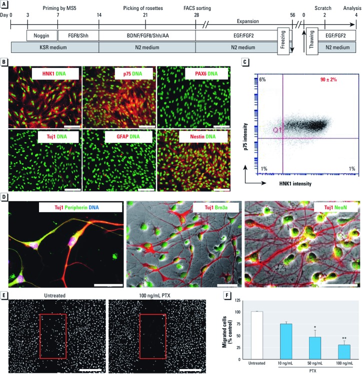
Methods: We generated NC cells from human embryonic stem cells, and after establishing a migration assay of NC cells (MINC assay), we tested environmental toxicants as well as inhibitors of physiological signal transduction pathways.

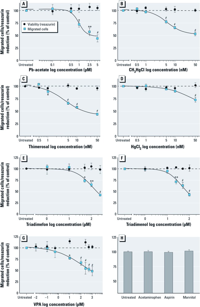
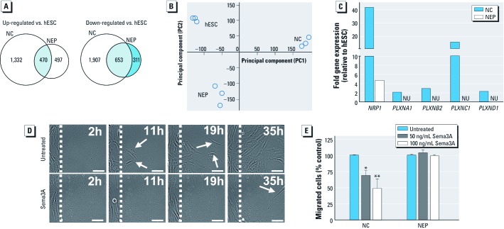
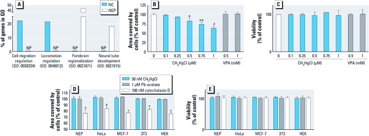
Results: Methylmercury (50 nM), valproic acid (> 10 µM), and lead-acetate [Pb(CH3CO2)4] (1 µM) affected the migration of NC cells more potently than migration of other cell types. The MINC assay correctly identified the NC toxicants triadimefon and triadimenol. Additionally, it showed different sensitivities to various organic and inorganic mercury compounds. Using the MINC assay and applying classic pharmacologic inhibitors and large-scale microarray gene expression profiling, we found several signaling pathways that are relevant for the migration of NC cells.

Conclusions: The MINC assay faithfully models human NC cell migration, and it reveals impairment of this function by developmental toxicants with good sensitivity and specificity.


Conflict of interest statement

The authors declare they have no actual or potential competing financial interests.

Figures

Figure 1
Figure 1
Characterization of hESC-derived NC cells. The schematic representation (A) illustrates the differentiation protocol and experimental procedures; AA, ascorbic acid; BDNF, brain-derived neurotrophic factor; FACS, fluorescence-activated cell sorting; FGF8, fibroblast growth factor-2; MS5, type of stromal cells; noggin, bone morphogenic protein antagonist; Shh, sonic hedgehog. (B) Immunocytochemical characterization of hESC-derived NC cells after thawing; bars = 200 µm; labels are color-keyed to images. (C) Flow cytometry analysis of NC cells for HNK1 and p75 expression. (D) Immunofluorescence analysis of peripheral neurons differentiated from NC cells; bars = 50 µm ; labels are color-keyed to images. (E) Representative images of cell migration in the absence or presence of pertussis toxin (PTX); bars = 500 µm. (F) Quantification of NC cell migration in the presence of PTX. The viability of the cells, as tested by resazurin reduction was 100 ± 5% under all conditions. *p < 0.05, and ** p < 0.01 compared with untreated controls.
Figure 2
Figure 2
Inhibition of cell migration in NC cells by heavy metals and known NC toxicants shown by the effects of different compounds on NC cell migration and viability (resazurin assay). (A) Pb(CH3CO2)4. (B) CH3HgCl. (C) Thimerosal. (D) HgCl2. (E) Triadimefon. (F) Triadimenol. (G) VPA. (H) High concentrations (250 µM) of acetaminophen, aspirin, or mannitol did not alter NC cell migration. Data are presented as mean ± SD of at least three independent experiments normalized to untreated controls. * p < 0.05, ** p < 0.01, and # p < 0.001 compared with untreated controls.
Figure 3
Figure 3
Transcriptome analysis of NC cells and specific migratory control. (A) Genome-wide transcription profiles were obtained for hESC, NC cells, and NEP cells; the number of significantly up-regulated or down-regulated genes is shown for NC cells and NEP cells relative to hESC. (B) Two-dimensional principal component analysis of the chip data; each circle indicates one experiment (n = 3 for each cell type). (C) Comparison of semaphorin receptor (NRP1, neuropilin 1; PLXN, plexin) expression in NC cells and NEP cells (NU, not up-regulated). (D) Migration of untreated or semaphorin 3A-exposed (Sema3A; 100 ng/mL) NC cells was recorded by video microscopy [see Supplemental Material, videos S1 and S2 (http://dx.doi.org/10.1289/ehp.1104489)]. Representative images are shown for four time points; white arrows indicate migrating cells; bars = 50 µm. (E) Quantification of NC cell and NEP cell migration in the presence of Sema3A.
Figure 4
Figure 4
Migration capacity and its modulation in different cell types. (A) Examples for gene ontologies with strongly differential expression in NC cells and NEP cells. The four chosen GOs contained on average 170 genes. The fraction of genes identified to be up-regulated is indicated; NP, not present. (B,C) Effect of increasing concentrations of CH3HgCl and VPA on NEP cell migration and cell viability. (D,E) Quantification of cell migration and cell viability of four different cell lines and NEP cells in the presence of MeHg, Pb(CH3CO2)4, and cytochalasin D; data are normalized to respective untreated controls (set to 100%) and presented as mean ± SD of three independent experiments. * p < 0.05, ** p < 0.01, and # p < 0.001 compared with untreated controls.

Comment in

References

    1. Andersen ME, Clewell HJ, III, Carmichael PL, Boekelheide K.2011Can case study approaches speed implementation of the NRC report: “Toxicity Testing in the 21st Century: A Vision and a Strategy?” Altex 283175–182. - PMC - PubMed
    1. Chambers SM, Fasano CA, Papapetrou EP, Tomishima M, Sadelain M, Studer L. Highly efficient neural conversion of human ES and iPS cells by dual inhibition of SMAD signaling. Nat Biotechnol. 2009;27(3):275–280. - PMC - PubMed
    1. Cowden J, Padnos B, Hunter D, MacPhail R, Jensen K, Padilla S. Developmental exposure to valproate and ethanol alters locomotor activity and retino-tectal projection area in zebrafish embryos. Reprod Toxicol. 2012;33(2):165–173. - PubMed
    1. Crofton KM, Mundy WR, Lein PJ, Bal-Price A, Coecke S, Seiler AE, et al. Developmental neurotoxicity testing: recommendations for developing alternative methods for the screening and prioritization of chemicals. Altex. 2011;28(1):9–15. - PubMed
    1. Di Renzo F, Broccia ML, Giavini E, Menegola E. Antifungal triazole derivative triadimefon induces ectopic maxillary cartilage by altering the morphogenesis of the first branchial arch. Birth Defects Res B Dev Reprod Toxicol. 2007;80(1):2–11. - PubMed

Publication types

LinkOut - more resources