Comparative Study
. 2012 Oct 7;9(75):2563-80.
doi: 10.1098/rsif.2012.0191. Epub 2012 May 9.

Structure and optical function of amorphous photonic nanostructures from avian feather barbs: a comparative small angle X-ray scattering (SAXS) analysis of 230 bird species

Affiliations
Comparative Study

Structure and optical function of amorphous photonic nanostructures from avian feather barbs: a comparative small angle X-ray scattering (SAXS) analysis of 230 bird species

Vinodkumar Saranathan et al. J R Soc Interface. .

Abstract

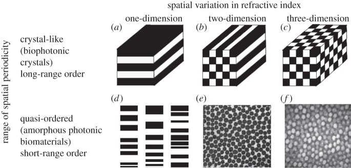
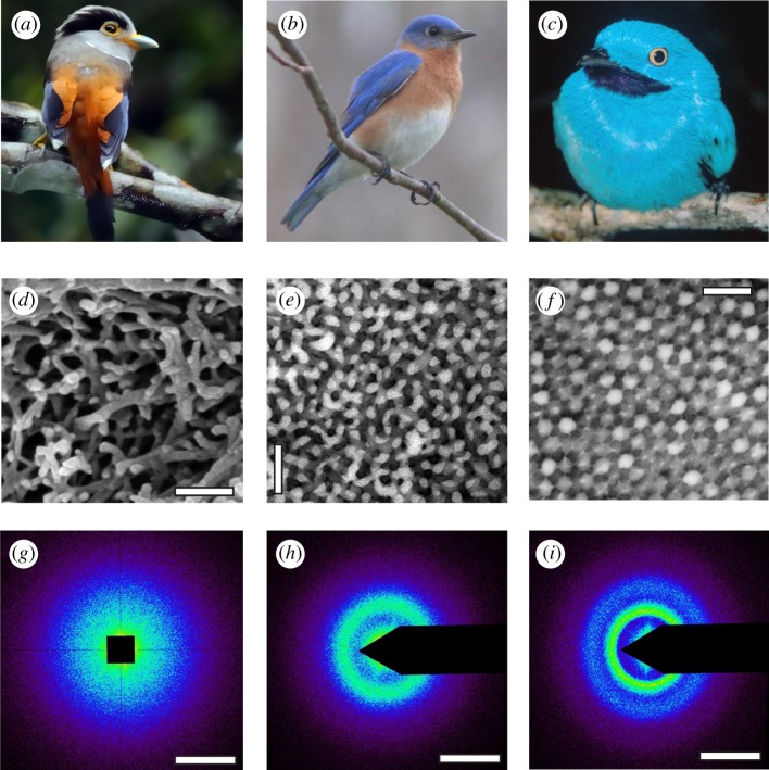
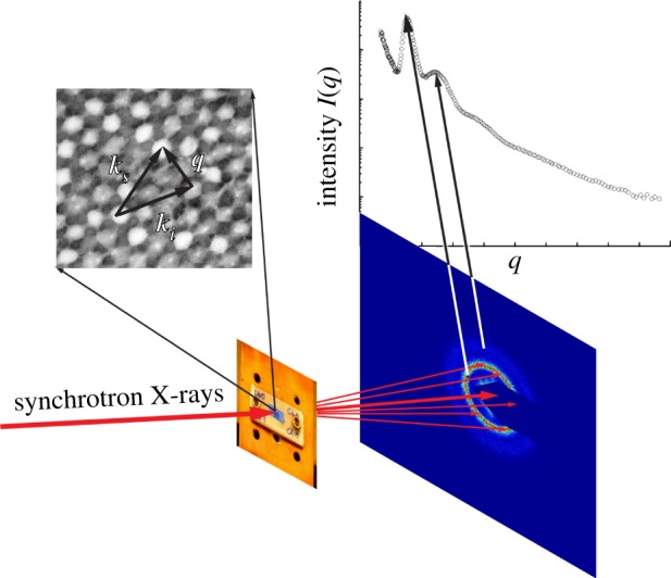
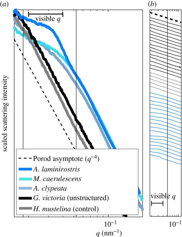
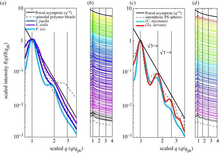
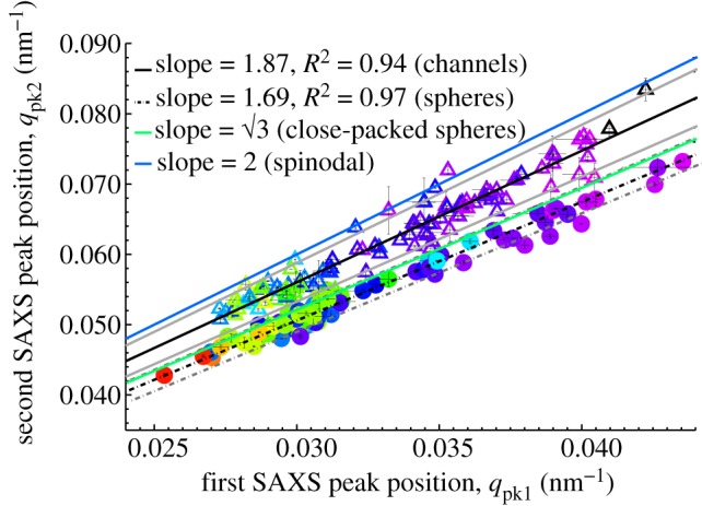
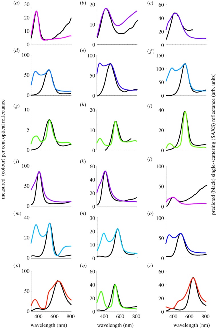
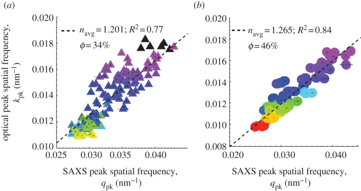
Non-iridescent structural colours of feathers are a diverse and an important part of the phenotype of many birds. These colours are generally produced by three-dimensional, amorphous (or quasi-ordered) spongy β-keratin and air nanostructures found in the medullary cells of feather barbs. Two main classes of three-dimensional barb nanostructures are known, characterized by a tortuous network of air channels or a close packing of spheroidal air cavities. Using synchrotron small angle X-ray scattering (SAXS) and optical spectrophotometry, we characterized the nanostructure and optical function of 297 distinctly coloured feathers from 230 species belonging to 163 genera in 51 avian families. The SAXS data provided quantitative diagnoses of the channel- and sphere-type nanostructures, and confirmed the presence of a predominant, isotropic length scale of variation in refractive index that produces strong reinforcement of a narrow band of scattered wavelengths. The SAXS structural data identified a new class of rudimentary or weakly nanostructured feathers responsible for slate-grey, and blue-grey structural colours. SAXS structural data provided good predictions of the single-scattering peak of the optical reflectance of the feathers. The SAXS structural measurements of channel- and sphere-type nanostructures are also similar to experimental scattering data from synthetic soft matter systems that self-assemble by phase separation. These results further support the hypothesis that colour-producing protein and air nanostructures in feather barbs are probably self-assembled by arrested phase separation of polymerizing β-keratin from the cytoplasm of medullary cells. Such avian amorphous photonic nanostructures with isotropic optical properties may provide biomimetic inspiration for photonic technology.


Figures

Figure 1.
Figure 1.
A classification of biophotonic nanostructural diversity based on the dimensionality of spatial variation in refractive index and its range of periodicity. (ac) One-, two- and three-dimensional biological photonic crystals with long-range periodic order in refractive index modulations (after Joannopoulos [6]). (d) A chirped lamellar stack, a one-dimensional quasi-ordered nanostructure with short-range spatial periodicity (currently unknown in birds; after Parker [2]). (e) TEM cross section of a two-dimensional amorphous or quasi-ordered nanostructure with short-range order comprising of parallel collagen fibres in a mucopolysaccharide matrix from the green tongue of magnificent bird-of-paradise (Cicinnurus magnificus, Paradisaeidae). (f) TEM cross section of a three-dimensional amorphous or quasi-ordered nanostructure of β-keratin and spheroidal air vacuoles from the spongy medullary cells of the azure blue crown feather barbs of male Blue-crowned Manakin (Lepidothrix coronata, Pipridae) with short-range quasi-periodic order.
Figure 2.
Figure 2.
Diversity of non-iridescent feather barb structural colours in birds and morphology of their underlying three-dimensional amorphous photonic nanostructures with short-range quasi-periodic order. (a) Female Silver-breasted Broadbill (Serilophus lunatus, Eurylaimidae). (b) Male Eastern Bluebird (S. sialis, Turdidae). (c) Male Plum-throated Cotinga (Cotinga maynana, Cotingidae). (d) SEM image of a rudimentary nanostructure with a very thin layer (1 μm or less) of a disordered network of spongy β-keratin bars present at the periphery of the medullary barb cells from the pale blue-grey primary coverts of S. lunatus, (e) TEM image of a channel-type β-keratin and air nanostructure from royal blue back contour feather barbs of S. sialis. (f) TEM image of a sphere-type β-keratin and air nanostructure from the dark turquoise blue back contour feather barbs of C. maynana. (gi) Representative two-dimensional small-angle X-ray scattering (SAXS) diffraction patterns for the rudimentary, channel- and sphere-type feather barb nanostructures in (df), respectively. The SAXS patterns for both channel- and sphere-type nanostructures exhibit ring-like features that demonstrate the isotropy and short-range spatial periodicity of these nanostructures, whereas the rudimentary barb nanostructure shows a diffuse, disc-like pattern. The false colour encoding corresponds to the logarithm of the X-ray scattering intensity. Scale bars: (d) 250 nm; (e,f) 500 nm; (gi) 0.05 nm−1. Photo credits: (a) Yiwen Yiwen (image in the public domain); (b) Ken Thomas (image in the public domain); and (c) Thomas Valqui (reproduced with permission).
Figure 3.
Figure 3.
Experimental schematic for SAXS experiments on feather barb nanostructures. A small (approx. 50 mm2) sample of the distal pennaceous portion of the feather vane is shown affixed to cover a 3 mm diameter hole on an aluminium block, which is then mounted in a plane perpendicular to the incident X-ray beam. The two-dimensional SAXS diffraction patterns for both channel- and sphere-type nanostructures exhibit ring-like features. Exploiting the circular symmetry of the SAXS diffraction patterns, the scattering intensity (I) is azimuthally averaged as a function of q to obtain scattering profiles, where the peaks correspond to the rings observed in the respective two-dimensional diffraction patterns. The scattering wavevector q measures the momentum transfer or the magnitude and direction of the scattering of incident photons (ki into ks) as a result of constructive interference from structural correlations of size 2π/q within the nanostructure.
Figure 4.
Figure 4.
SAXS structural diagnosis of weakly structured, control and unstructured feather barbs. (a) Representative azimuthal SAXS profiles for the rudimentary sphere-type nanostructure (‘structured*’, electronic supplementary material, table S2) in A. laminirostris (Ramphastidae), and the rudimentary channel-type nanostructures (‘structured’, see electronic supplementary material, table S2) in Melanotis caerulescens (Mimidae) and Anas clypeata (Anatidae) as well as unstructured feather barbs from Goura victoria (Columbidae) and Hylocichla mustelina (Turdidae). The azimuthal profiles are normalized to one along the intensity axis for ease of comparison. (b) The azimuthal SAXS profiles for 18 weakly structured (blue lines), five control (grey lines) and 16 unstructured feather barbs (black lines) on a semi-log scale. The azimuthal profiles are vertically displaced along the intensity axis for clarity. The azimuthal scattering profiles of the control feathers, many purple, magenta and bright white feathers as well as several marginally blue-grey (black lines) feathers did not deviate from Porod's Law even at low q (<0.04 nm−1). Thus, these feathers do not possess any underlying barb nanostructure, ruling out any contribution of constructive interference to their observed colours. The azimuthal SAXS profiles from feathers with mainly slaty blue-black to pale greyish-blue colours show slight to moderate deviations from Porod's Law at low q, with these features resembling a shoulder rather than a peak. Nevertheless, the spatial correlations that these feather barbs do possess appear to be at the appropriate length scales to be able to produce visible structural colours through interference. (a,b) The thick horizontal line indicates the range of spatial frequencies relevant for avian visible structural colour production.
Figure 5.
Figure 5.
SAXS structural diagnosis of amorphous photonic nanostructures in feather barbs. (a) and (c) depict, respectively, representative normalized azimuthal SAXS profiles for channel-type nanostructures from I. puella (Irenidae), S. sialis (Turdidae), and Pitta iris (Pittidae) and sphere-type nanostructures from C. maynana (Cotingidae) and Tangara larvata (Thraupidae) on a log–log scale exhibiting clearly distinguishable structural differences. The azimuthal profiles are scaled to compare across different colours and nanostructural sizes. (b) and (d) show, respectively, scaled azimuthal SAXS profiles for 159 channel- and 96 sphere-type nanostructures on a semi-log scale. The colour of each profile is coded to the approximate colour of the corresponding feathers based on its primary optical peak hue (pure UV colours shown in black). These azimuthal profiles are vertically displaced along the y-axis for clarity. In addition to the primary peak, the channel-type nanostructures (a,b) either have a weak to a pronounced shoulder at approximately twice the dominant spatial frequency, 2*qpk or lack any other significant feature, while the sphere-type nanostructures (c,d) exhibit one or more pronounced higher-order scattering peaks in addition to the primary peak at ratios of approximately √3 and √7 times qpk. The grey dashed lines in all figures plot the scaled experimental scattering profiles from two polymer mixtures undergoing spinodal decomposition [33,34] (a,b) and an amorphous film of self-assembled colloidal polymer spheres (c,d). The vertical lines at 1,2,3 (a,b) and at 1,√3 and √7 (c,d) are visual guides for the expected positional ratios for the SAXS peaks based on experimental observations of classical spinodal and nucleated, close-packed sphere morphologies, respectively.
Figure 6.
Figure 6.
Regression plot of the first and second SAXS peaks of channel- (open triangles) and sphere-type (shaded circles) amorphous barb nanostructures. The colour of each triangle or circle is coded to the approximate colour of the corresponding feather (UV colours in black). The thin vertical and horizontal lines at each data point indicate the standard error of the mean (s.e.m). The solid blue and green lines with corresponding slopes of 2 and √3 indicate the expected positional ratios for the second SAXS peak based on experimental observations of spinodal and nucleated, close-packed sphere morphologies, respectively. The solid and dashed grey lines, respectively, indicate the 95% confidence interval of the regressions.
Figure 7.
Figure 7.
Single-scattering SAXS reflectance predictions for the primary optical peaks of channel (ai) and sphere-type (jr) amorphous barb nanostructures. SAXS single-scattering reflectance predictions (black lines) and measured normal incidence reflectance curves (coloured lines) for (a) UV (black) belly feather barbs of Charmosyna papou (Psittacidae), (b) violet primary feather barbs of Acryllium vulturinum (Psittacidae), (c) royal blue rump feather barbs of S. sialis (Turdidae), (d) sky blue rump feather barbs of Alcedo atthis (Alcedinidae), (e) deep azure blue back feather barbs of Irena puella (Irenidae), (f) electric blue wing covert feather barbs of Pitta maxima (Pittidae), (g) emerald green back feather barbs of Ailuroedus buccoides (Ptilonorhynchidae), (h) emerald green back feather barbs of Charmosyna papou (Psittacidae), (i) emerald green back feather barbs of Calyptomena whitehadi (Eurylaimidae), (j) deep blue throat feather barbs of Tangara chilensis (Thraupidae), (k) royal blue wing covert feather barbs of Wetmorethraupis sterrhopteron (Thraupidae), (l) violet scapular feather barbs of Conirostrum albifrons (Thraupidae), (m) dark turquoise blue back feather barbs of C. maynana (Cotingidae), (n) sky blue back feather barbs of male Tersina viridis (Thraupidae), (o) azure blue rump feather barbs of Lepidothrix serena (Pipridae), (p) golden yellow crown feather barbs of Lepidothrix vilasboasi (Pipridae), (q) electric green back feather barbs of Chloronis riefferii (Thraupidae), (r) golden crown feather barbs of Tangara larvata (Thraupidae). The colour of the measured reflectance curves is approximately coded to the colour of the feather barbs based on the spectral position of the primary reflectance peak.
Figure 8.
Figure 8.
Regression plots of the primary optical peak hue from normal incidence reflectance measurements expressed as peak spatial frequency (kpk = 2π/λpk) against the dominant spatial frequency of structural correlations (qpk) measured using SAXS for (a) channel- (shaded triangles) and (b) sphere-type (shaded circles) nanostructures. For both nanostructural classes, the size of the nanostructural periodicity measured by SAXS strongly predicts, i.e. scales with the measured primary peak hue, demonstrating that the underlying barb nanostructures are tuned to produce the observed structural colours. The inverse of twice the slope of the regression yields navg, the average or effective refractive index (and hence ϕ, the keratin volume fraction) for each class of nanostructure. The estimated navg and φ for sphere nanostructures on the whole (1.265, 46%) is significantly higher than that for channel morphologies (1.201, 34%) and congruent with predictions of the phase separation hypothesis. The colour of each triangle or circle is coded to the approximate colour of the corresponding feather (UV colours in black). The vertical and horizontal lines at each data point indicate the standard error of the mean (s.e.m).

References

    1. Kinoshita S., Yoshioka S., Miyazaki J. 2008. Physics of structural colors. Rep. Prog. Phys. 71, 076401.10.1088/0034-4885/71/7/076401 (doi:10.1088/0034-4885/71/7/076401) - DOI - DOI
    1. Parker A. R. 2000. 515 million years of structural colour. J. Opt. A Pure Appl. Opt. 2, R15–R2810.1088/1464-4258/2/6/201 (doi:10.1088/1464-4258/2/6/201) - DOI - DOI
    1. Prum R. O. 2006. Anatomy, physics, and evolution of avian structural colors. In Bird coloration, volume 1 mechanisms and measurements (eds Hill G. E., McGraw K. J.), pp. 295–353 Cambridge, MA: Harvard University Press
    1. Srinivasarao M. 1999. Nano-optics in the biological world: beetles, butterflies, birds, and moths. Chem. Rev. 99, 1935–196110.1021/cr970080y (doi:10.1021/cr970080y) - DOI - DOI - PubMed
    1. Vukusic P., Sambles J. R. 2003. Photonic structures in biology. Nature 424, 852–85510.1038/nature01941 (doi:10.1038/nature01941) - DOI - DOI - PubMed

Publication types

LinkOut - more resources